Biomedical visualization: LIVE at the ROM

Pliosaur sketch by Ruth Chang. Photo credit: R. Chang

Pliosaur sketch by Ruth Chang. Photo credit: R. Chang

From May to June, and again from October to November, the 103-year-old Royal Ontario Museumhosts “Friday Night Live.” ROM Friday Night Live transforms the museum from a venue for art, culture and natural history into one of the most desirable social destinations in Toronto. Patrons dressed in cocktail attire form a line outside and along Bloor Street to purchase one of the small number of tickets available at the door for the sold-out event. The venerable old institution lowers the lights and fills with the beat of dance music played by DJs. Guests enjoy drinks and explore offerings from pop-up food vendors while experiencing live performances and special demonstrations in the ROM’s gallery exhibits.

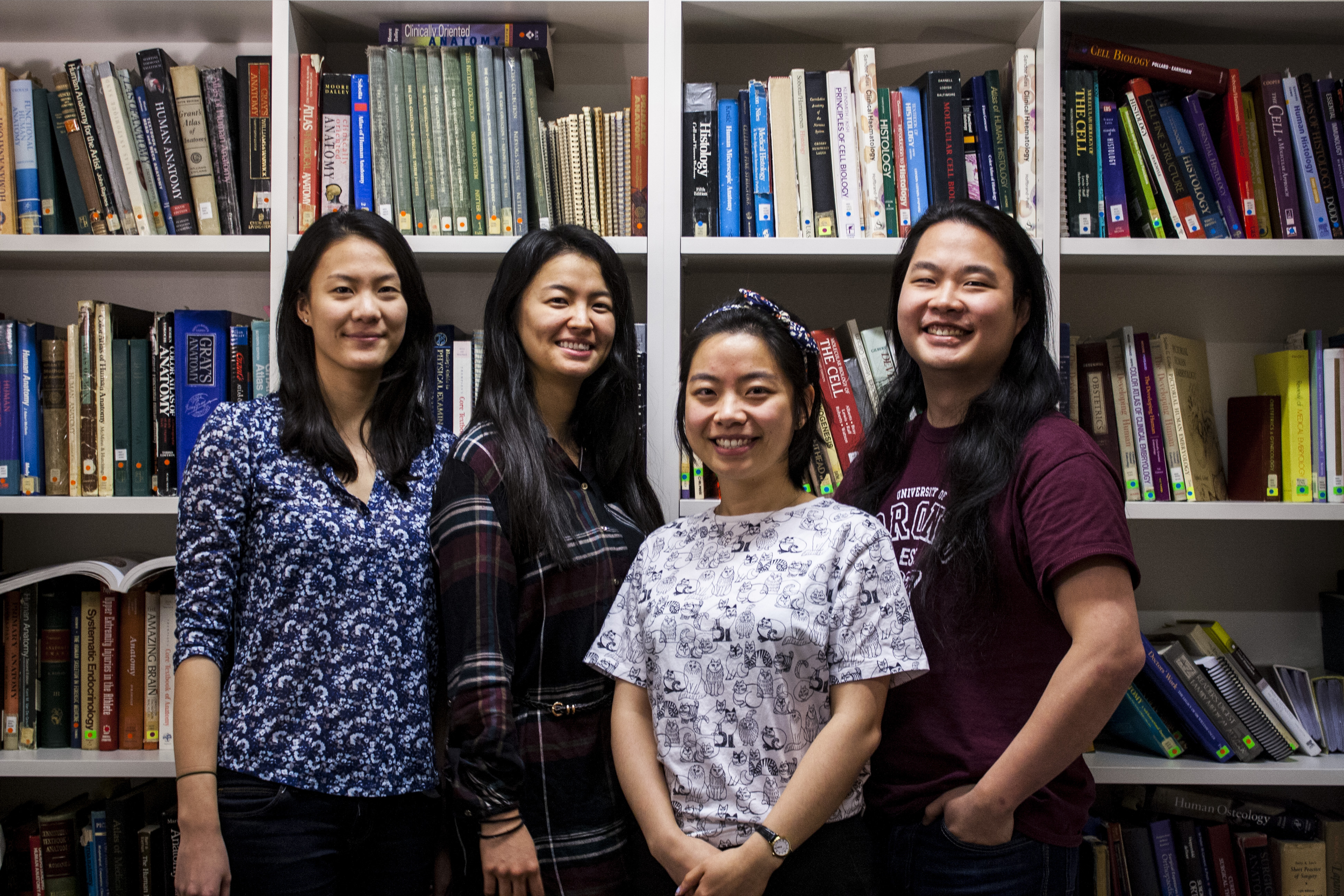
The ROM’s festival programs organizers invited four students from the Master of Science in Biomedical Communications to give a live demonstration of scientific visualization at the November 18, 2016 production of ROM Friday Night Live.

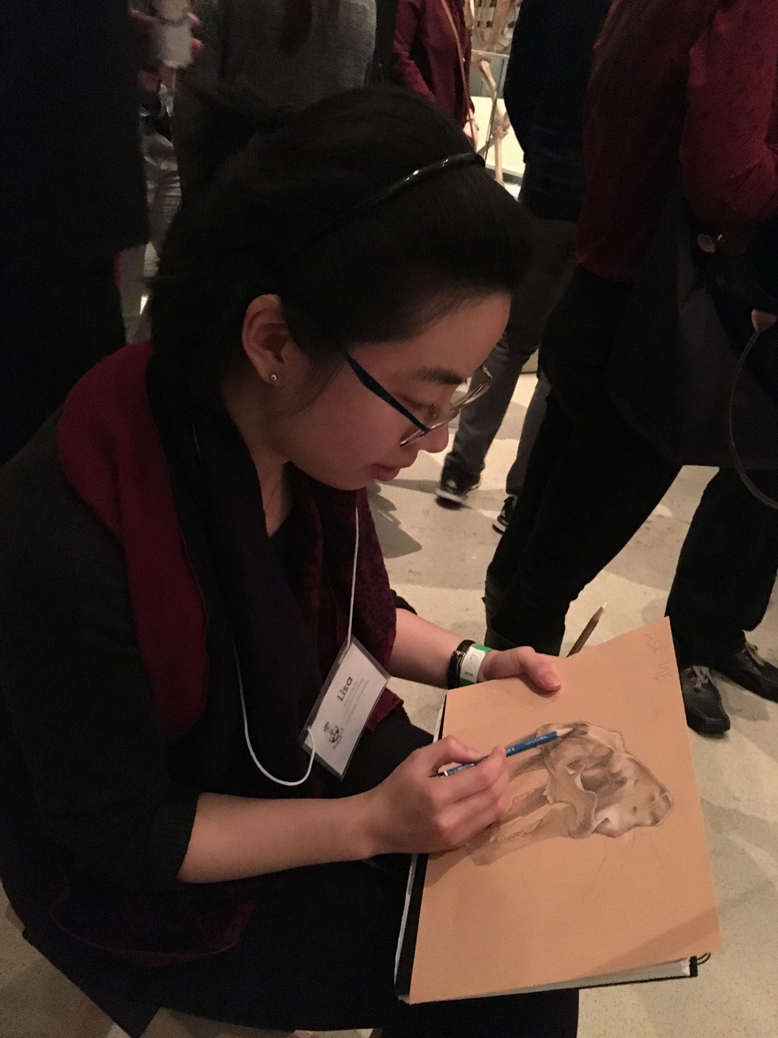
The volunteers, first year students Nancy JiRyan Park and Lisa Qiu, and second year student Ruth Chang, attended an orientation at the museum the Tuesday before the event. They were shown the galleries where they would draw and each student received a pair of event tickets to share with friends.

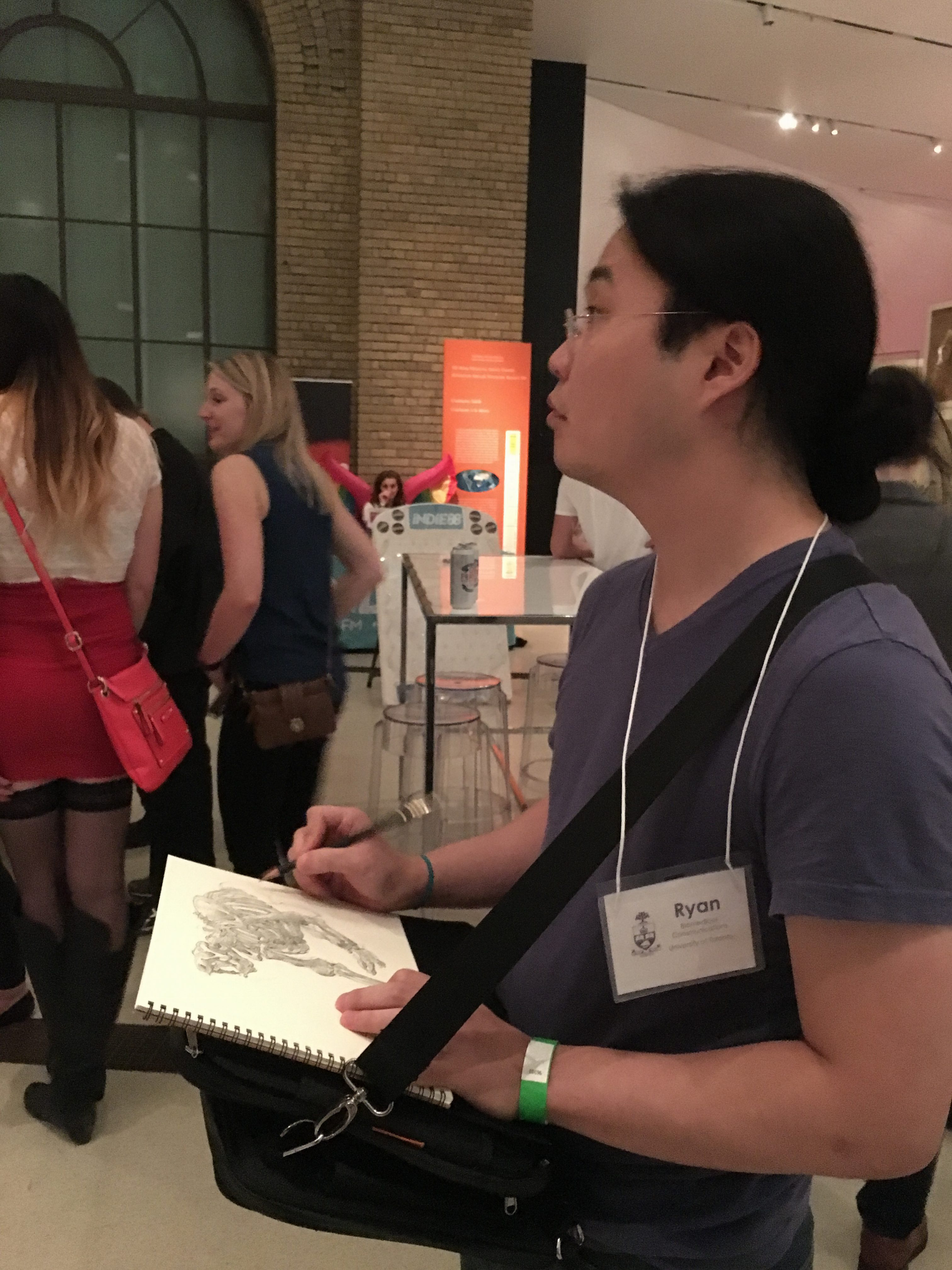
On the night of the event, the students were stationed in the James and Louise Temerty Galleries of the Age of the Dinosaurs and the Reed Gallery of the Age of Mammals but they were free to move between and within the galleries.

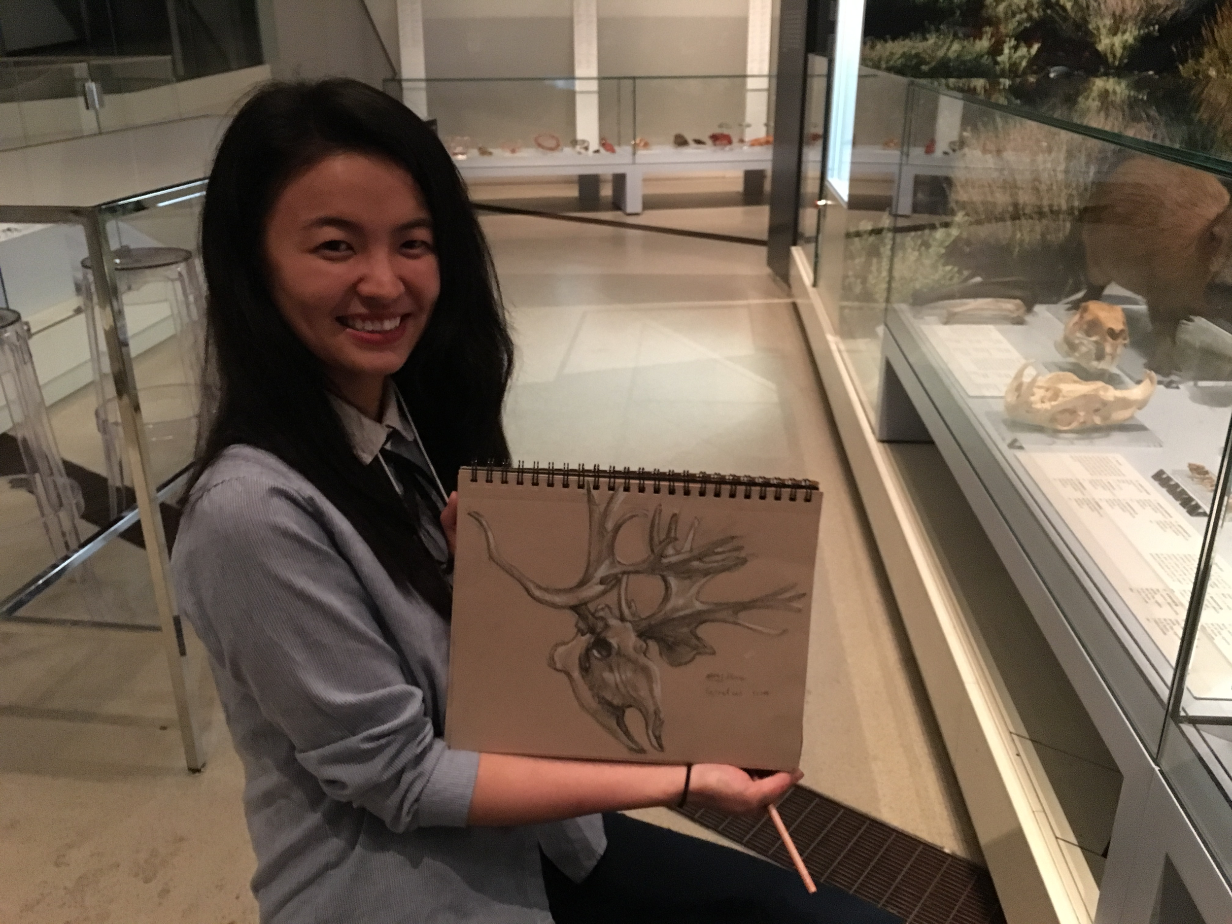
“At least one student migrated into the Gallery of the Birds,” says Jessica Hawthorn, one of the festival coordinators and former UTM biology graduate student.

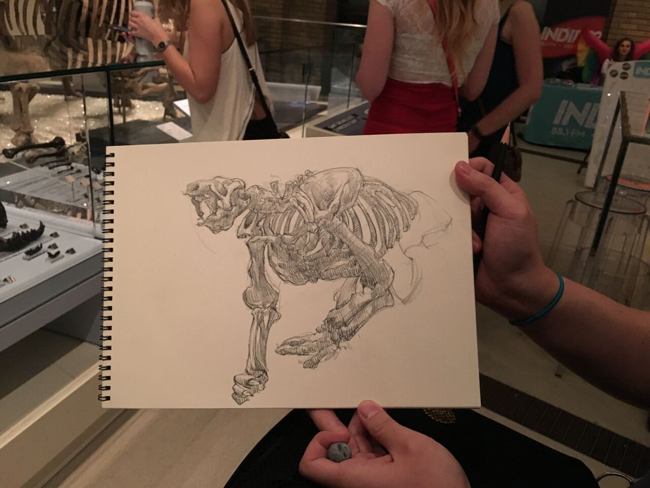
At first, party-goers were hesitant to interrupt the students as they sat or stood to sketch. Ryan Park swung his name tag around and wore it on his back while he drew the skeleton of the ROM’s giant ground sloth. “Then people were comfortable addressing me by name,” he says. Park estimates that he spoke to about 20 small groups of two or three people throughout the night.

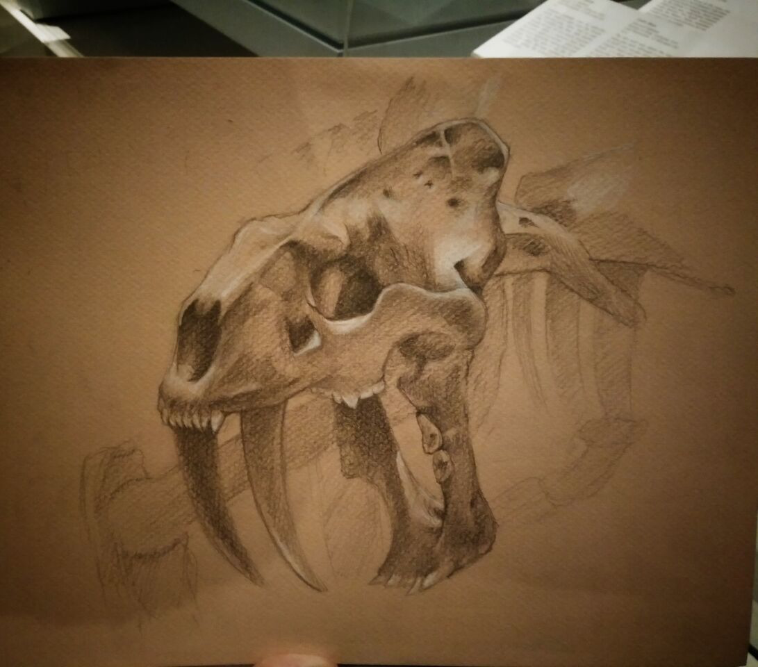
Ruth Chang wrote “ask me questions” on her sketchpad above her sketch of a pliosaur. “People stopped hovering and started approaching me freely after that,” she says.

Festival coordinator Hawthorn says that including biomedical communications students in ROM Friday Night Live highlights how illustration is a medium for communicating scientific concepts and how the museum is a library of objects. The museum is a repository of specimens that hold information on evolution and ecological change and these specimens can be accessed by researchers and illustrators.

Biomedical communications students regularly access the museum’s collections for their research. In one instance, Chi-Chun Liu, who worked with UTM biologist Sanja Hinić-Frlog and biomedical communications professor David Mazierski, was exploring the biomechanics of an extinct flightless bird for a scientific animation. ROM curators took a specimen off exhibit and gave Liu exclusive access for his direct observation and measurement.

“That is the great value of our collection,” says Hawthorn.

Now finished its fifth year and tenth full season, the motivation behind ROM Friday Night Live was to engage that twenty-something demographic. “We created a night that takes everything we do from exhibitions to galleries to research and wove it through a program where food and entertainment are factored in,” says Chris Kennedy, the ROM’s senior manager of festival programs.

ROM Friday Night Live typically sells out two to three weekends in advance and has an average attendance of 3,000 people. The night the biomedical communications students gave their live demonstration a remarkable 3,400 guests attended. ROM Friday Night Live has become an important source for revenue that supports the ROM’s operations, collections and research, says Kennedy. “But most importantly, it encourages people to get in and explore all our gallery exhibitions.”

To brighten and warm the cold, bleak month of February, ROM festival organizers offer a one-off Friday Night Live. This year on February 3, 2017, “Afro Fête” kicks of the ROM’s Black History Month programming. ROM Friday Night Live’s all-new spring programming premieres May 5.

by Maeve Doyle

Stephen Gilbert Award 2016

Savanna Jackson, 2016 recipient of Stephen Gilbert award

Savanna Jackson, 2016 recipient of Stephen Gilbert award

Second year Biomedical Communications graduate student Savanna Jackson was named the 2016 recipient of the Professor Stephen Gilbert Award for Excellence in Biomedical Visualization. Program director Nicholas Woolridge made the announcement Friday, December 16, 2016 after the annual Year End Critique. A committee of Biomedical Communications faculty selected Jackson’s piece Atherosclerosis from 42 submissions made by first and second year students in the program.

Jackson, who holds a bachelor of science in biology and a certificate in art and design, joined the Biomedical Communications program in fall 2015. She created Atherosclerosis for the course MSC2018H Visual Representation of Processes in Pathology taught by Professor Shelley Wall.

Jackson said that her goal in creating the piece was to capture atherosclerosis on both the cellular and gross anatomical level. “I wanted to demonstrate the highly complex cellular composition of an atherosclerotic plaque while also clearly exhibiting how the slow buildup of local cellular changes within a plaque can have large scale effects on cardiovascular health.” She applied a minimalist aesthetic to the text and background elements in order to provide contrast to the illustration’s intricacies.

“Savanna’s piece shows a classic pathological change in a tissue landscape over time and with an elegant integration of pictorial information and graphic design elements,” said Woolridge. “It is an excellent example of very clear illustrative storytelling.”

“When I saw my piece appear on the screen behind Nick, I’d describe my feelings as pleasantly shocked and incredibly honoured,” said Jackson.

Currently, Jackson is creating a 2D animation to explain her Master’s Research Project CANVIS: Citation, Annotation & Visualization Integration System, a web application that will allow medical illustrators to embed citation information directly into their visual media.

The Biomedical Communications program established the Professor Stephen Gilbert Award in memory of Professor Gilbert who passed away in February 2014. Gilbert, who taught part-time for 23 years in the Biomedical Communications/Art as Applied to Medicine program at the University of Toronto, was an internationally recognized medical illustrator and master of the pen-and-ink technique.

by Maeve Doyle

BMC graduate student fundraiser 2016

BMC Holiday Card Fundraiser 2016

BMC Holiday Card Fundraiser 2016

Biomedical Communications students and faculty donated all-original illustrations to create a set of 17 holiday-themed cards. Available for $20 per set, the authentic BMC-made cards sold out in a day which prompted a second run. In just three days this December, the students raised over $700 for CP24’s CHUM Christmas Wish. The Christmas Wish provides toy and financial support to ~300,000 people in need across the greater Toronto area.

Presentation of the inaugural Nancy Grahame Joy Award

MScBMC Year I student Lisa Qiu, Nancy Grahame Joy’s niece Dorothy Lothian, Director Nicholas Woolridge (Photo cred: Marc Dryer)

MScBMC Year I student Lisa Qiu, Nancy Grahame Joy’s niece Dorothy Lothian, Director Nicholas Woolridge (Photo cred: Marc Dryer)

Nancy Grahame Joy’s niece presented the inaugural Professor Nancy Grahame Joy Biomedical Communications Entrance Award to first-year Biomedical Communications student Lisa Qiu. Dorothy Lothian made the presentation Tuesday, November 8, 2016 at the Class of 2016 Final Presentations. The Nancy Grahame Joy Award recognizes an exceptional applicant to the program upon their registration in the first year of studies.

Qiu graduated from the University of Toronto in 2016 with an Honours Bachelor of Science, specialising in pathobiology. She received numerous awards of merit during her undergraduate tenure and was twice named a Dean’s List Scholar. She also pursued formal art training, part-time, at The Academy of Realist Art.

Professor Joy, one of the illustrators of Grant’s Atlas of Anatomy, was chair of the Department of Art as Applied to Medicine (now Biomedical Communications) for 23 years. The Nancy Grahame Joy Award was made possible through a bequest from Professor Joy, who passed away in 2013.

“We are really glad to see how this has developed,” said Lothian, who attended with other members of the Joy family.

Each year near convocation, the BMC program invites the students in the graduating class to present their capstone biomedical visualization projects to their peers, families and friends.

“It is really interesting to see what the students are producing 30 years after Nancy was head of the department,” said Lothian.

At the final presentations, awardees from the graduating Class of 2016 were recognized with the presentation of certificates by program director Nicholas Woolridge.

Melissa Phachanhla received the Eila I. Ross Memorial Scholarship for demonstrating excellence and professionalism throughout her tenure in the program.

Wendy Gu, who also holds the first-ever Stephen Gilbert Award for Artistic Excellence, received the inaugural Sherwood P. and Judith Gebhard Smith Award.

Samantha HolmesSarah Kim and Susan Le received the Wendy M. Kates Memorial Award in recognition of the academic achievement they demonstrated through their Master’s Research Projects.

The Class of 2016 formally joined the family of BMC alumni the following day, November 9, at Fall Convocation.

 by Maeve Doyle

Gifts of support to the Biomedical Communications program can be made through “Giving to BMC.”

BMC student wraps up summer with creation of a web app

Matan (1T7) Berson spent the summer of 2016 working at the Toronto Video Atlas for Surgery (TVASurg). Berson collaborated with the TVASurg team on the creation of an interactive web application that allows users to manipulate and explore the 3D anatomic models shown in the Video Atlas.

Now in the second year of the MScBMC program, Berson intends to apply the technical and design skills he developed on this project to his master’s research project and he hopes for future opportunities to create interactive applications for medical education on mobile, web and virtual reality platforms.

TVASurg team members include MScBMC alumni Albert (1T0) Fung, Paul (1T1) Kelly and Joy (1T3) Qu. Part of Perioperative Interactive Education (PIE) at the Toronto General Hospital, TVASurg creates training tools for surgeons, which combine surgical video with animation.

You can read more about Berson’s interactive web application on TVASurg’s summer wrap up blog post: http://pie.med.utoronto.ca/TVASurg/summer-wrap-up-featuring-matan-berson/

BMC graduate student explains the Zika virus through animation

When reports of the Zika virus outbreak first appeared in the media in 2015, the messages were uncertain, confusing and not intended for the people directly impacted by the virus. In response, Meriem Benlamri, a second year Biomedical Communications graduate student, created the 2D animation “What is the Zika virus?” She saw a need to communicate information about the virus effectively through a visual medium and to bring clarity to the epidemic specifically for the populations at greatest risk.

Benlamri, who holds an Honours Bachelor of Science in biology, began by researching the Zika virus, its link to microcephaly, and the mosquito that transmits the virus. Until April 2016, there was no scientific evidence that could confirm the link between the virus and microcephaly.

“Even the World Health Organization could only say that the link was suspected,” Benlamri says.

She found that the information people needed to protect themselves and to prevent the spread of the virus was available from public health organizations. But she also found that the information, for some audiences, was too textual and too medical. Existing visuals only incorporated video recordings of experts who spoke about the virus. Fact sheets often ended with travel advisories for those who live outside Central and South America, the sites of the outbreak. Benlamri knew that she could create an animation to explain the outbreak and engage her target audience long enough to give them the information they needed.

She wrote a script to explain what the virus is, how it is transmitted and where in the world the current outbreak is. She explains the symptoms of the virus in adults and the link between the Zika virus and microcephaly. She describes steps that the impacted population can take to protect themselves from contracting the virus. She wrote the script so that the narration could be visually represented at the same time.

Next, Benlamri built the 2D visual elements in vector-based software and brought the graphics into compositing software to animate them. Then, she recorded and optimized the audio and imported that into the compositing software. Finally, she exported all the content as a video.

Benlamri created the one-and-a-half minute, 2D animation as part of an exercise in storytelling for the biomedical communications graduate course “Advanced Media Design.”

“Meriem has created an animation in which all the information is significant. She presents it clearly and simply and her audience requires no specialized knowledge to understand it,” says Michael Corrin, Biomedical Communications faculty and the course’s instructor. “She makes the information memorable through visuals that reinforce what is being spoken and she engages the audience through her use of colour and rapidly moving imagery.”

On April 13, 2016, the Centers for Disease Control and Prevention published a media statement that confirmed Zika virus as a cause of microcephaly. Benlamri is revising her piece to reflect this confirmed link.

Although her animation is currently available only in English, to reach her target audiences, Benlamri is collaborating with a translator on a Spanish version of the script. She hopes to find another translator to create a Portuguese version and she has written to the Pan American Health Organization to discuss the distribution of her animation. Benlamri wants to get her animation to people in Central and South America and specifically to pregnant women because they and their babies are at the highest risk, she says.

“I was impressed by the passion Meriem brought to the project. Her investment in the topic, its importance and timeliness had a huge impact on what she produced,” says Corrin. “Her project could inform people both inside and outside the areas effected by the Zika virus outbreak.”

Next, Benlamri begins work on her master’s research project to develop web-based interactive modules that incorporate 2D animations and 3D models to educate psychiatry residents about the latest advances in neuroscientific research.

by Maeve Doyle

Neurosurgeon recognized with original biomedical art

An original watercolour-and-ink painting created by Stacey Krumholtz was awarded by the Journal of Neurosurgery publishing group to neurosurgeon Dr. Martin Weiss to recognize his 20 years of service as editor-in-chief of Neurosurgical Focus.

Krumholtz, who graduated with a Master of Science in Biomedical Communications in 2015, is the Biomedical Communications Specialist at the Arthur & Sonia Labatt Brain Tumour Research Centre at SickKids Hospital in Toronto.

Dr. James Rutka, director of the Brain Tumour Research Centre and editor-in-chief of the Journal of Neurosurgery, commissioned the painting of endoscopic transphenoidal surgery for presentation to Dr. Weiss who will continue as associate editor of Neurosurgical Focus.

“Art is so subjective that it can be a tricky gift to create for someone you don’t know,” says Krumholtz, “but when it works out and they like it—I think it’s a hard gift to beat!”

Krumholtz’s piece was presented to Dr. Weiss at the AANS annual meeting which took place April 30 to May 4, 2016 in Chicago.

by Maeve Doyle

Paleo Puzzles: Art exhibit of fossil reconstruction illustrations

The Biomedical Communications program invites members of the university community to view a display of paleo illustrations created by Diane Scott in the Department of Biology at the University of Toronto Mississauga (UTM). The exhibit was mounted for the recent meeting of the Canadian Society of Vertebrate Palaeontology held at UTM, May 19 to 21, 2016. The display includes more than 40 samples from a collection of palaeontological specimen and fossil reconstruction illustrations.

Scott, a lab technician, fossil preparator and scientific illustrator begins her work with material that to the untrained eye resembles a lump of rock. With the aid of a microscope and small tools, she removes as much of the rocky matrix as possible to reveal the tiny animal hidden there. If the fossil is a previously unknown specimen, she researches the literature for similar animals to help her distinguish the important bony landmarks from cracks in the fossil. She photographs the fossil, brings the photograph into digital art software and creates an outline. She prints the outline onto coquille board and, with the original specimen as reference, hand-shades the outline to give it form and texture.

“I just love doing the work. It’s like a puzzle for me. Assembling the pieces of a fossil is like preparing a sculpture,” says Scott.

Scott first demonstrated her talent as a scientific illustrator in 1979 as one of Robert Reisz’s undergraduate students and she has worked with the renowned vertebrate palaeontologist ever since. She has co-authored over 40 peer-reviewed papers and she teaches graduate and undergraduate students how to prepare and illustrate fossils.

“Because of the University of Toronto’s reputation, we receive pieces from around the world to prepare,” says Scott. She might remove more of the matrix to reveal more of the fossil or she might repair damage to the pieces. Fossils are often returned to their institutions in better condition than they were received and many have become museum exhibits. “I am blessed to work with so many pieces from so many places.”

David Mazierski, associate professor in the Biomedical Communications program, who curated the paleo exhibit has long-held an interest in the role of palaeontological illustration and its importance in depicting fossil evidence.

“I’ve always admired Diane’s work and it is, in fact, part of the reason I pursued my own studies in vertebrate palaeontology,” he says.

Mazierski worked with Scott to select illustrations from those stored in Professor Reisz’s lab. He rescued three of the oldest drawings which had been taped into the windows of a room in the Davis Building. He repaired the damaged pieces and scanned the collection to create a digital archive of the works to make them available for reproduction.

Next, he mounted and matted the art with archival quality materials and researched each piece to create information placards. These interpretive signs include the importance of the organism that was studied, a geological time scale to place the organism in time, and a citation for the publication in which the piece appeared.

“Going through the collection with Dave really made me appreciate how much I’ve been able to accomplish over the years,” says Scott.

Visitors can view the exhibit from Monday to Friday between 8:30 and 4:30 in the Biomedical Communications hallway on the third floor of the Health Sciences Complex at the University of Toronto Mississauga until September 30, 2016.

BMC grad students illustrate immunology publication

Top: Ruth Cheng, Lauren Huff; Bottom: Matan Berson, Ursula Florjanczyk

Top: Ruth Cheng, Lauren Huff; Bottom: Matan Berson, Ursula Florjanczyk

Matan Berson, Ruth Chang, Ursula Florjanczyk and Lauren Huff, first year graduate students in the Biomedical Communications (BMC) program, illustrated an article in the April 2016 issue of Trends in Molecular Medicine. They created the illustrations for the course “Visual Representation of Medical Knowledge” taught by BMC associate professor Michael Corrin.

In the first half of the course, “Intracellular Illustration,” students develop visual storytelling skills they began in the textbook illustration portion of “Human Anatomy”. For the first time, they employ colour in illustration and learn to conceptualize events that happen at the cellular and molecular scale.

Students work with content advisors from the Department of Immunology. Stephen K.H. Li, postdoctoral research fellow, and Alberto Martin, associate professor, proposed topics to illustrate for their review article, “Mismatch repair and colon cancer: Mechanisms and therapies explored.”

Figure 1. Etiologic factors of colorectal cancer (CRC) by Ursula Florjanczyk

Figure 1. Etiologic factors of colorectal cancer (CRC) by Ursula Florjanczyk

Ursula Florjanczyk created Figures 1 and 3. “Figure 1. Etiologic factors of colorectal cancer (CRC)” shows different factors that effect CRC. “Figure 3. Mechanism of mammalian MMR” shows the molecular mechanism of DNA mismatch repair.

Lauren Huff illustrated “Figure 2. Genetic mutations resulting from MMR deficiency contribute to CRC Development.” In this piece, Huff depicts the journey of a crypt stem cell in the colon as it migrates up the crypt. She shows a healthy cell capable of mismatch repair and a cell which lacks mismatch repair machinery and initiates a CRC.

Figure 2. Genetic mutations resulting from MMR deficiency contribute to CRC Development by Lauren Huff

Figure 2. Genetic mutations resulting from MMR deficiency contribute to CRC Development by Lauren Huff

Figure 3. Mechanism of mammalian MMR by Ursula Florjanczyk

Figure 3. Mechanism of mammalian MMR by Ursula Florjanczyk

Matan Berson illustrated “Figure 4. Effects of inflammation and oxidative damage in CRC.” Berson shows how gut bacteria can cause an inflammatory response, which can in turn trigger mutations in and expressions of genes associated with CRC.

In “Figure 5. Effects of butyrate produced by the gut microbiota in MMR-proficient and MMR-deficient CRC murine models,” Ruth Chang illustrates the “Butyrate Paradox” in which butyrate produced by gut microbiota can cause the growth of tumours, but high levels of butyrate injected directly to the gut can suppress the growth of these tumours.

Figure 4. Effects of inflammation and oxidative damage in CRC by Matan Berson

Figure 4. Effects of inflammation and oxidative damage in CRC by Matan Berson

Figure 5. Effects of butyrate produced by the gut microbiota in MMR-proficient and MMR-deficient CRC murine models by Ruth Chang

Figure 5. Effects of butyrate produced by the gut microbiota in MMR-proficient and MMR-deficient CRC murine models by Ruth Chang

In the second half of the course, “Surgical Illustration,” students advance from one-page storytelling to multipage designs. In year two, students continue to develop skills with static media and begin formal instruction in motion and interactive media.

by Maeve Doyle

Recent news from the Jenkinson Lab

Jodie Jenkinson and Vijay Shahani stand in graduation gowns at the University of Toronto.

The Royal Society of Chemistry’s Chemistry Education Research and Practice (CERP) journal published Vijay (1T5) Shahani and Jodie Jenkinson’s paper, “The efficacy of interactive analogical models in the instruction of bond energy curves in undergraduate chemistry.”

The authors researched two analogies to introduce students to the concept of potential energy wells. Pre- and post-tests of the analogies demonstrated how critical the importance of design in education is.

The research paper appears in CERP’s April 2016 issue.

ACCN, Canadian Chemical News magazine published a science communications story about Naveen (1T5) Devasagayam and Jodie Jenkinson’s research into visualizing molecular interactions while capturing the relationships “between molecular structure, composition and spatial orientation within the tight confines of a living cell.”

The article, “Bringing an artist’s sensibility to molecular landscapes” by Tim Lougheed appears in the March/April 2016 issue of the magazine.

by Maeve Doyle