by Maeve Doyle
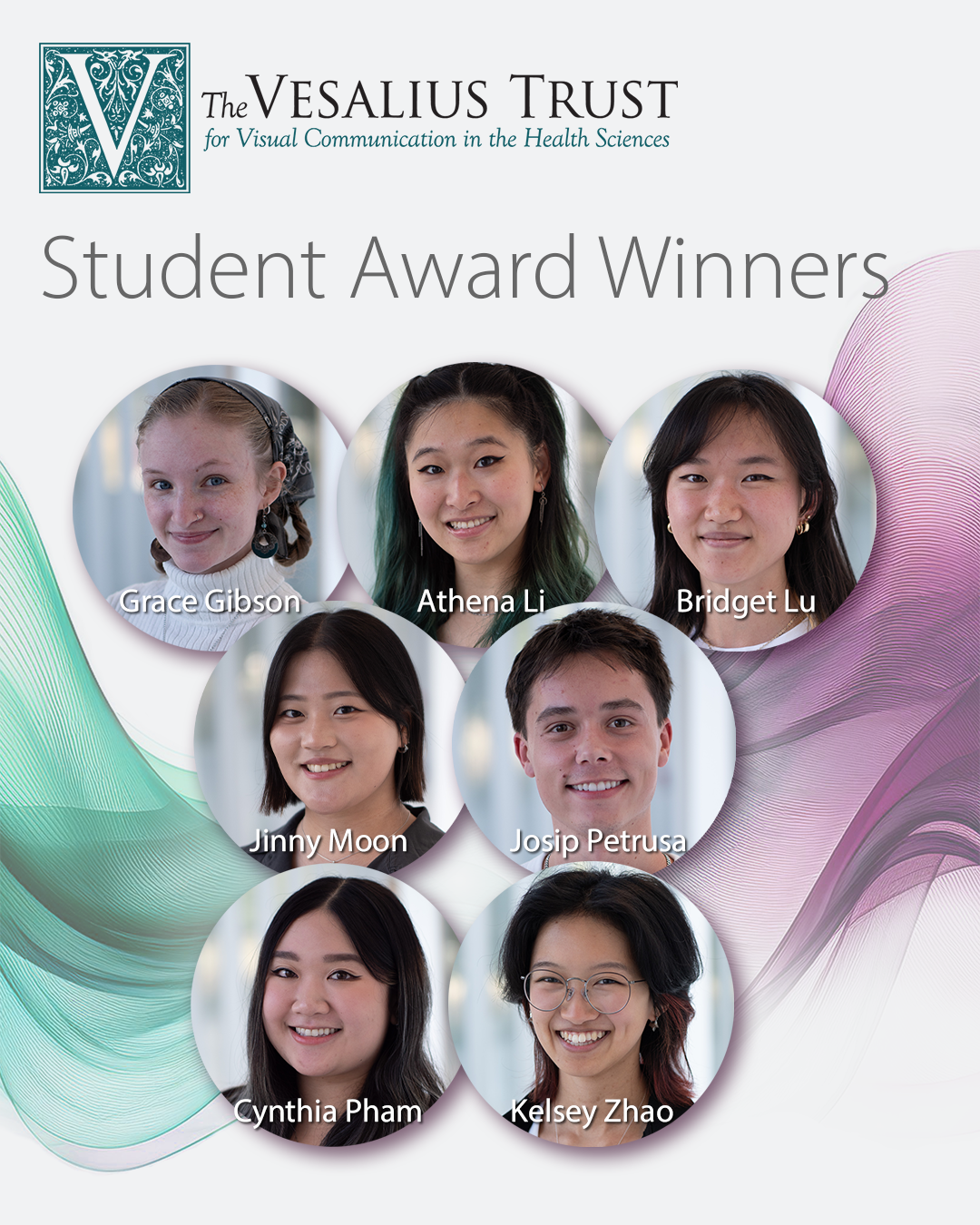
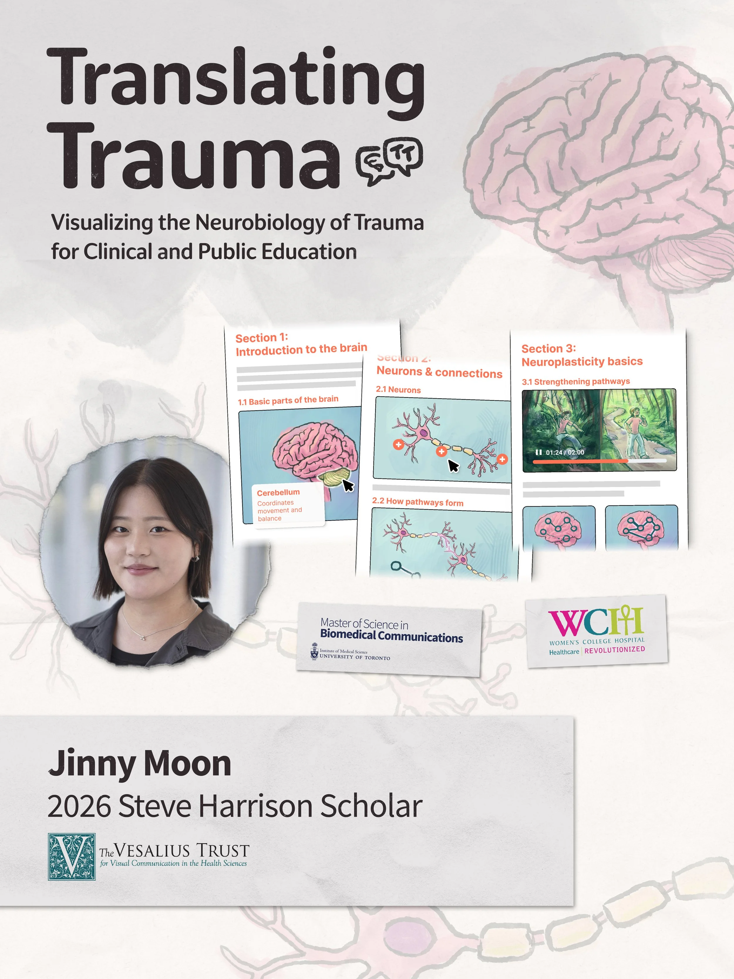
The Vesalius Trust named Jinny Moon the recipient of the 2026 Steve Harrison Award. The award recognizes and supports her research into visualizing the neurobiology of trauma.
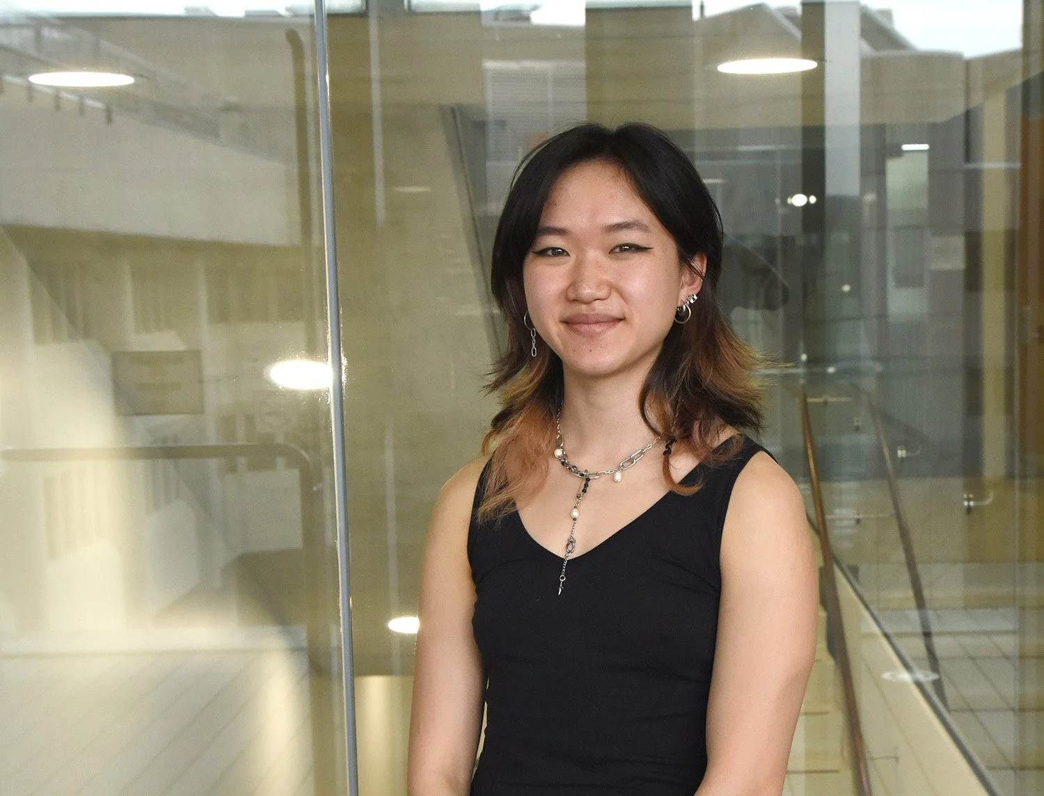
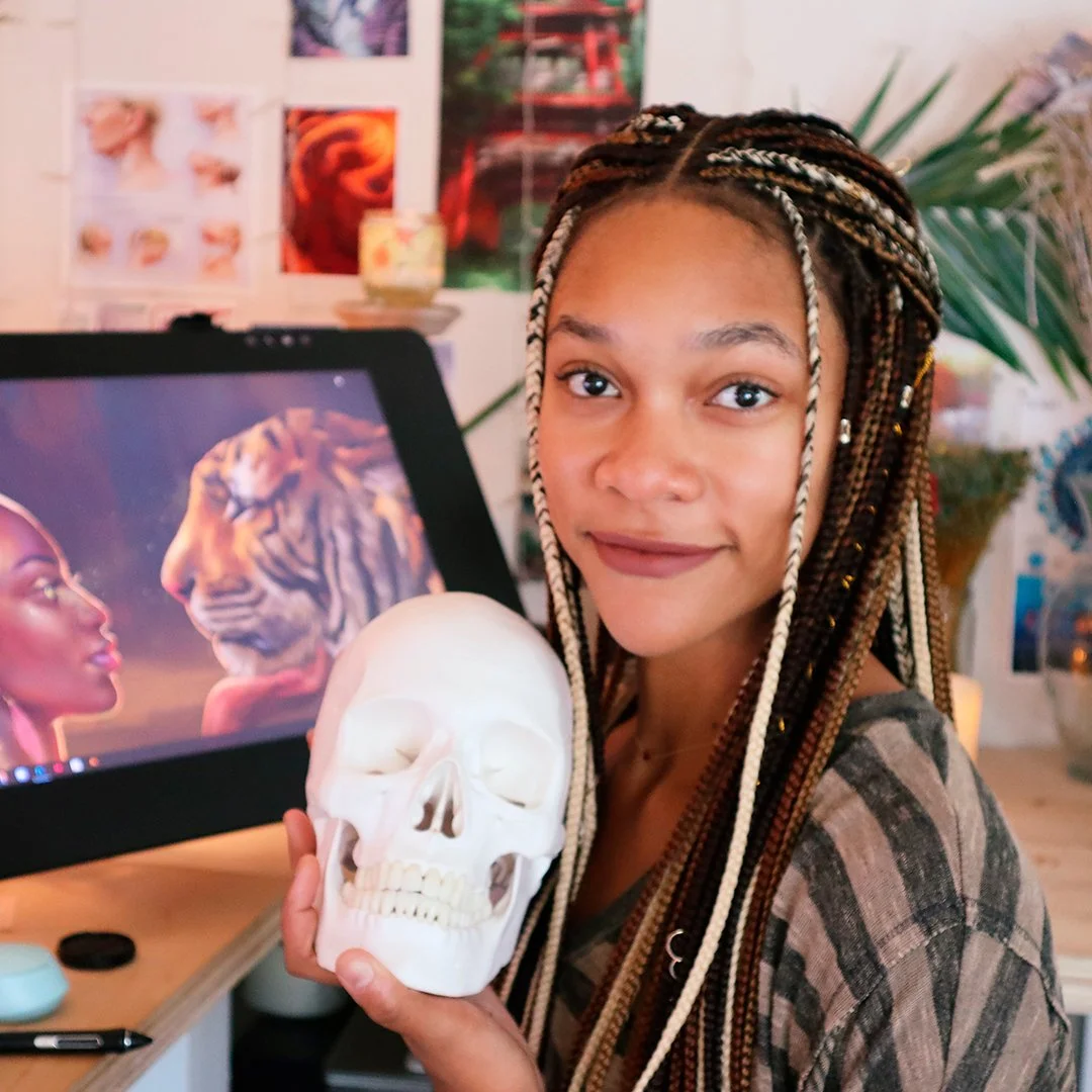
Moon is a second-year student in the Master of Science in Biomedical Communications at the University of Toronto. She received the award for her research proposal, Translating trauma: visualizing the neurobiology of trauma for clinical and public education.
She is conducting her research under the guidance of Dr. Dana Ross who is the co-director of the Trauma Therapy Network of Ontario at Women's College Hospital in Toronto. The program focuses on expanding access to trauma-focused care and supporting both patients and healthcare providers in understanding the impacts of trauma and pathways to healing.
"Trauma has profound effects on the brain and body, yet the underlying neuroscience can be difficult to translate into everyday clinical and lived experience," says Ross. Her goal is to have this science translated into accessible, visually engaging media that improve understanding of trauma and its effects.
Moon was immediately drawn to the project.
"I've always been interested in neurobiology, human behaviour, and cognition," she says. "I've also had a long-standing interest in public health and in mental health."
Her review of existing trauma education materials found several gaps: outdated science, inconsistent visuals and a lack of technical polish. Resources were either too complicated, too simple or reinforced stigma. Few addressed neuroplasticity as a way to explain both the effects of trauma and the brain's capacity to recover.
Moon is applying visual design principles to develop a user-centred online education module. It includes still images and a 2D animation that incorporates a visual metaphor to show how new practices and habits can help build and shape new response pathways.
"If the hospital's platform can support it, there will also be interactive diagrams with clickable hotspots," she says.
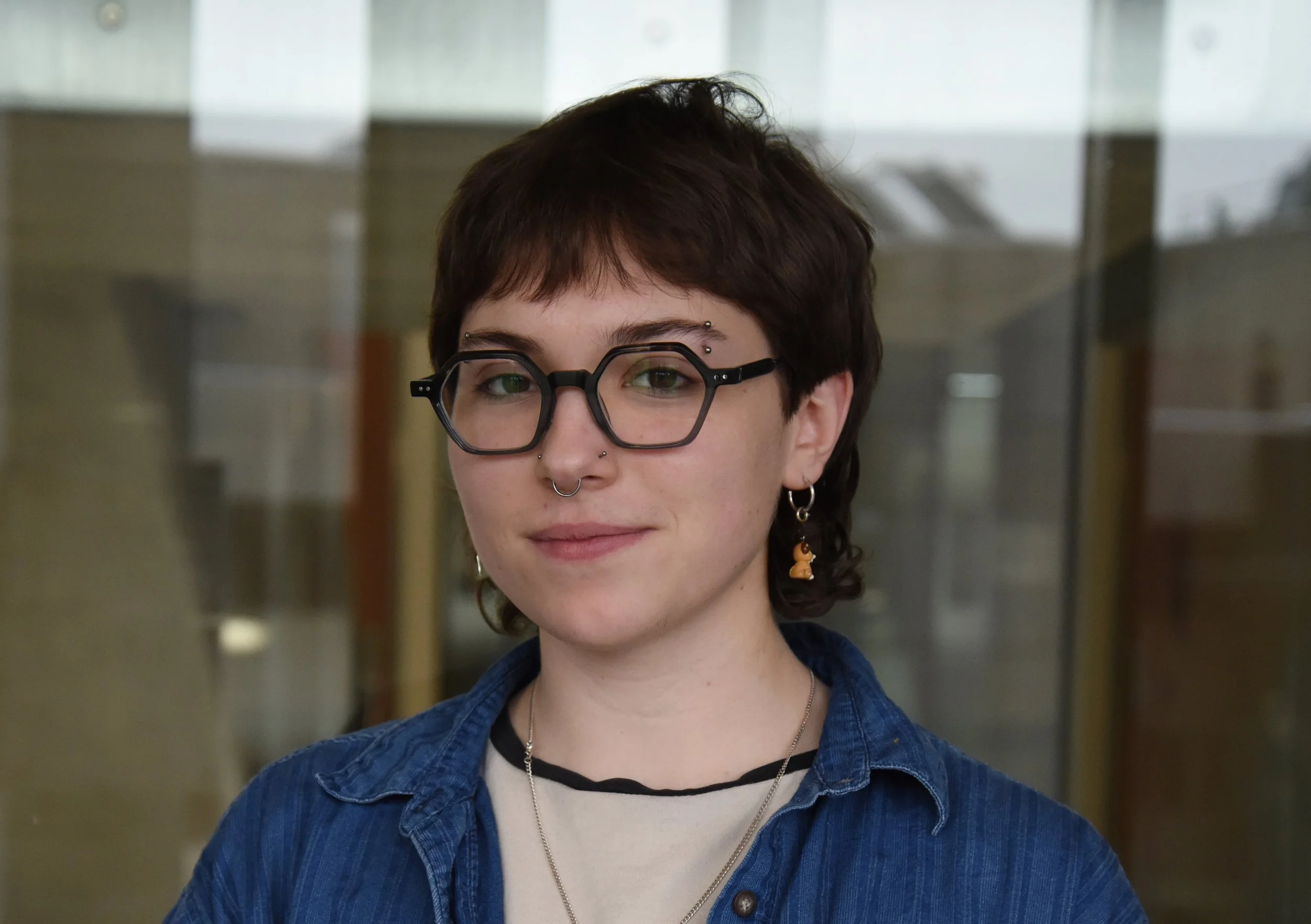
"Jinny is producing work that is both scientifically rigorous and deeply considered for its audience," says Professor Shehyrar Saharan, Moon’s supervisor. "I'm really happy that Jinny won. She really deserved it."
Moon says she felt both validated and humbled when she learned that she had received the Steve Harrison Award. "It was encouraging to know others recognized how important the work is and its impact for the audience it serves."
Saharan says that Moon is also preparing a detailed, annotated pictorial account of her process for publication. "This will be a valuable contribution to the biomedical visualization community," he says.
Moon continues to refine the animation, illustrations and narrative elements of her project to create a cohesive learning experience.
She hopes to see the work's integration into the Women's College Hospital's e-learning platform Trauma PORTAL for patients and their CARE training programs for healthcare professionals.
~
Web sites referenced
Jinny Moon’s online portfolio https://www.jinnymoon.ca
Announcement: Vesalius Trust 2026 student award winners https://bmc.med.utoronto.ca/news-events/2026/4/2/vesalius-trust-2026-student-award-winners
Women’s College Hospital’s Trauma Portal https://www.womenscollegehospital.ca/care-programs/mental-health/trauma-therapy-program/
Women’s College Hospital’s CARE training programs https://www.womenscollegehospital.ca/care-programs/mental-health/trauma-therapy-program/