

Top: Ruth Cheng, Lauren Huff; Bottom: Matan Berson, Ursula Florjanczyk
Matan Berson, Ruth Chang, Ursula Florjanczyk and Lauren Huff, first year graduate students in the Biomedical Communications (BMC) program, illustrated an article in the April 2016 issue of Trends in Molecular Medicine. They created the illustrations for the course “Visual Representation of Medical Knowledge” taught by BMC associate professor Michael Corrin.
In the first half of the course, “Intracellular Illustration,” students develop visual storytelling skills they began in the textbook illustration portion of “Human Anatomy”. For the first time, they employ colour in illustration and learn to conceptualize events that happen at the cellular and molecular scale.
Students work with content advisors from the Department of Immunology. Stephen K.H. Li, postdoctoral research fellow, and Alberto Martin, associate professor, proposed topics to illustrate for their review article, “Mismatch repair and colon cancer: Mechanisms and therapies explored.”

Figure 1. Etiologic factors of colorectal cancer (CRC) by Ursula Florjanczyk
Ursula Florjanczyk created Figures 1 and 3. “Figure 1. Etiologic factors of colorectal cancer (CRC)” shows different factors that effect CRC. “Figure 3. Mechanism of mammalian MMR” shows the molecular mechanism of DNA mismatch repair.
Lauren Huff illustrated “Figure 2. Genetic mutations resulting from MMR deficiency contribute to CRC Development.” In this piece, Huff depicts the journey of a crypt stem cell in the colon as it migrates up the crypt. She shows a healthy cell capable of mismatch repair and a cell which lacks mismatch repair machinery and initiates a CRC.
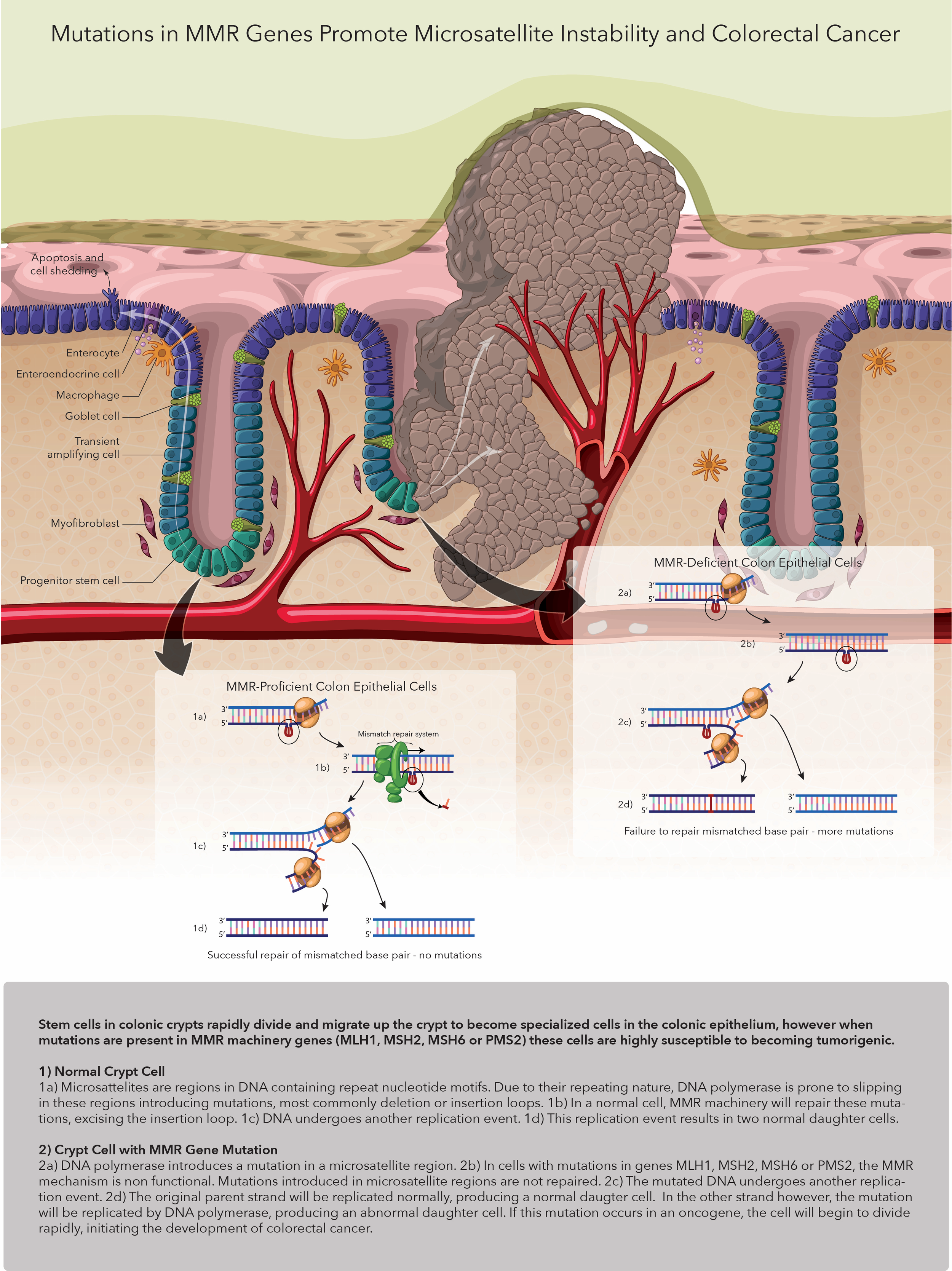
Figure 2. Genetic mutations resulting from MMR deficiency contribute to CRC Development by Lauren Huff
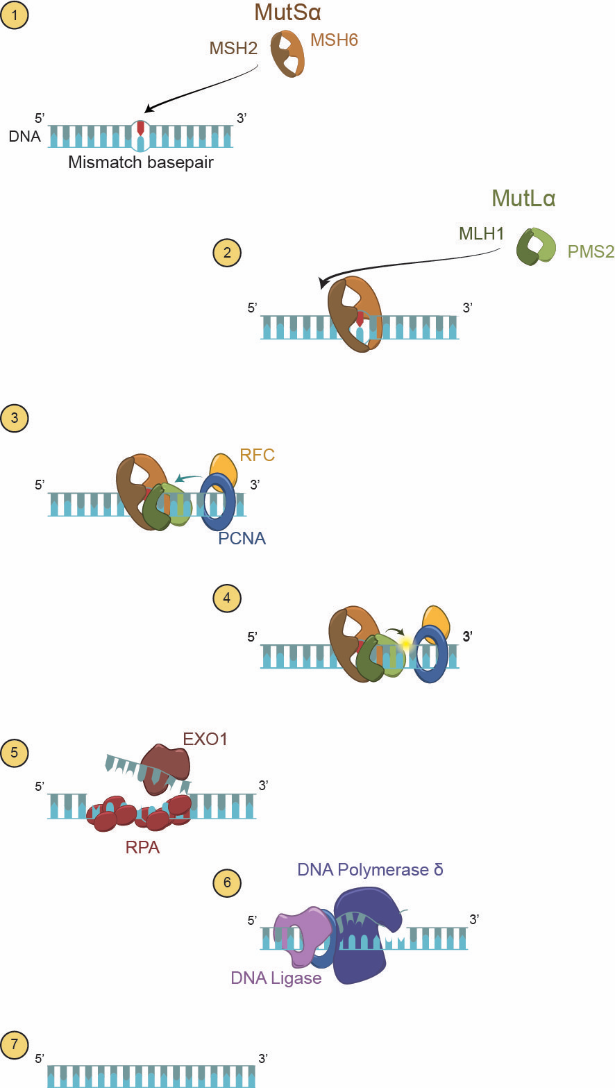
Figure 3. Mechanism of mammalian MMR by Ursula Florjanczyk
Matan Berson illustrated “Figure 4. Effects of inflammation and oxidative damage in CRC.” Berson shows how gut bacteria can cause an inflammatory response, which can in turn trigger mutations in and expressions of genes associated with CRC.
In “Figure 5. Effects of butyrate produced by the gut microbiota in MMR-proficient and MMR-deficient CRC murine models,” Ruth Chang illustrates the “Butyrate Paradox” in which butyrate produced by gut microbiota can cause the growth of tumours, but high levels of butyrate injected directly to the gut can suppress the growth of these tumours.
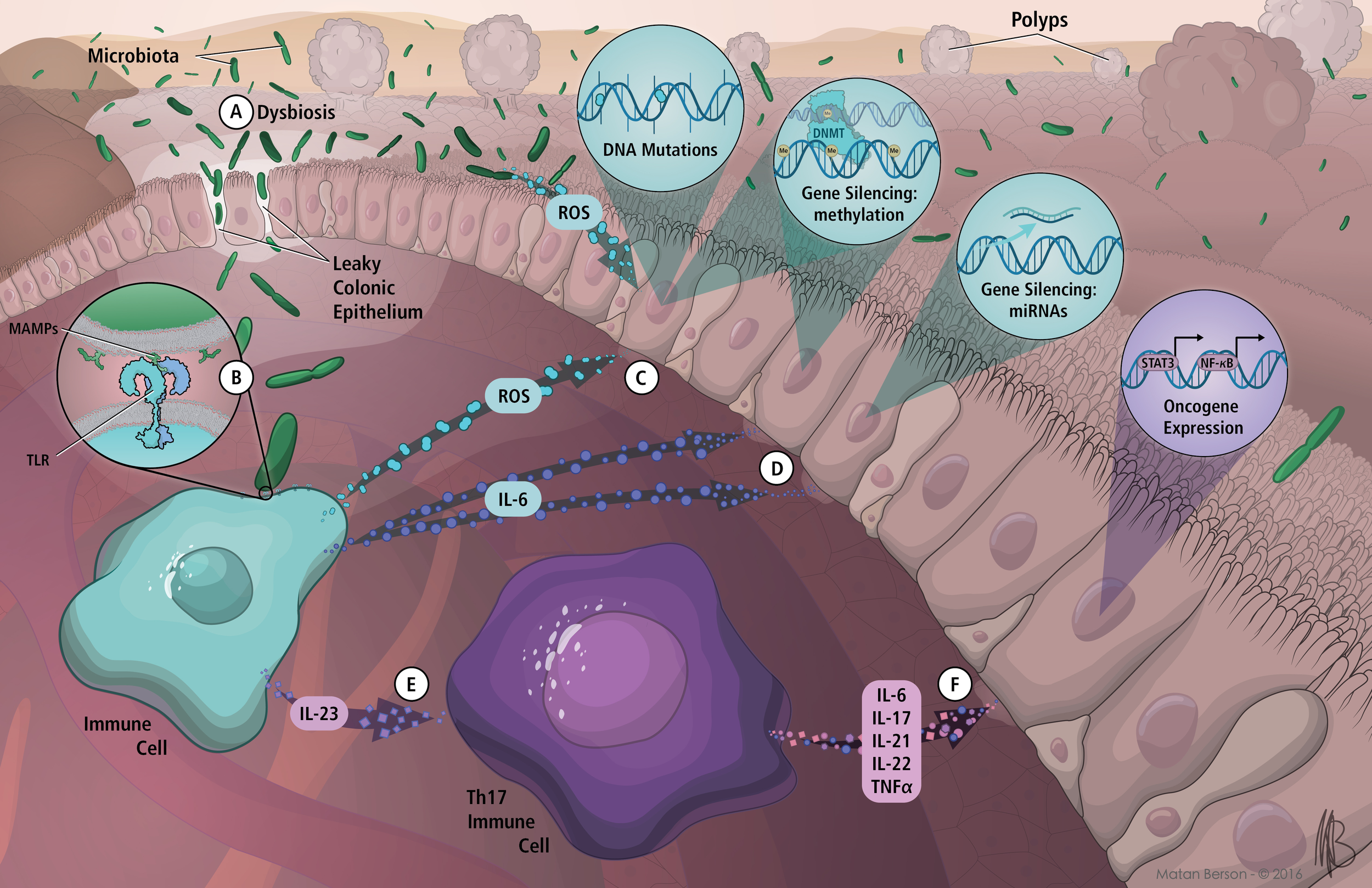
Figure 4. Effects of inflammation and oxidative damage in CRC by Matan Berson

Figure 5. Effects of butyrate produced by the gut microbiota in MMR-proficient and MMR-deficient CRC murine models by Ruth Chang
In the second half of the course, “Surgical Illustration,” students advance from one-page storytelling to multipage designs. In year two, students continue to develop skills with static media and begin formal instruction in motion and interactive media.
by Maeve Doyle