Spring Critique 2024

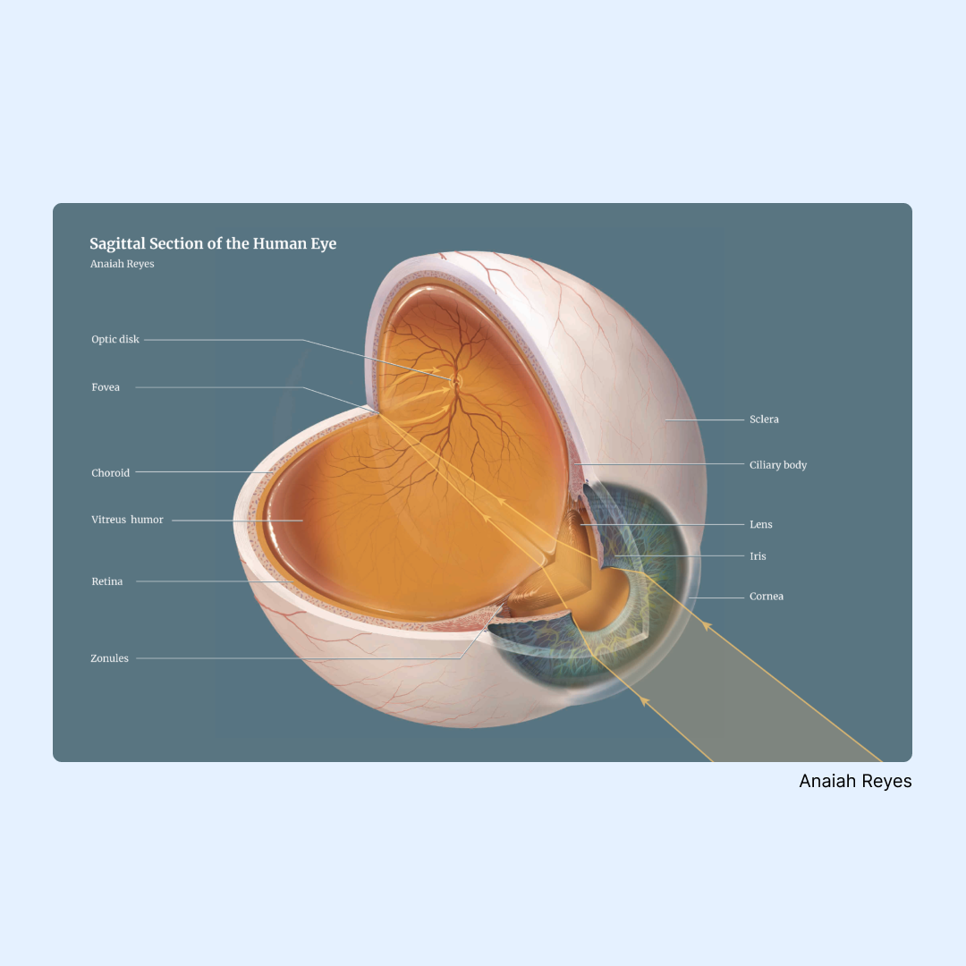
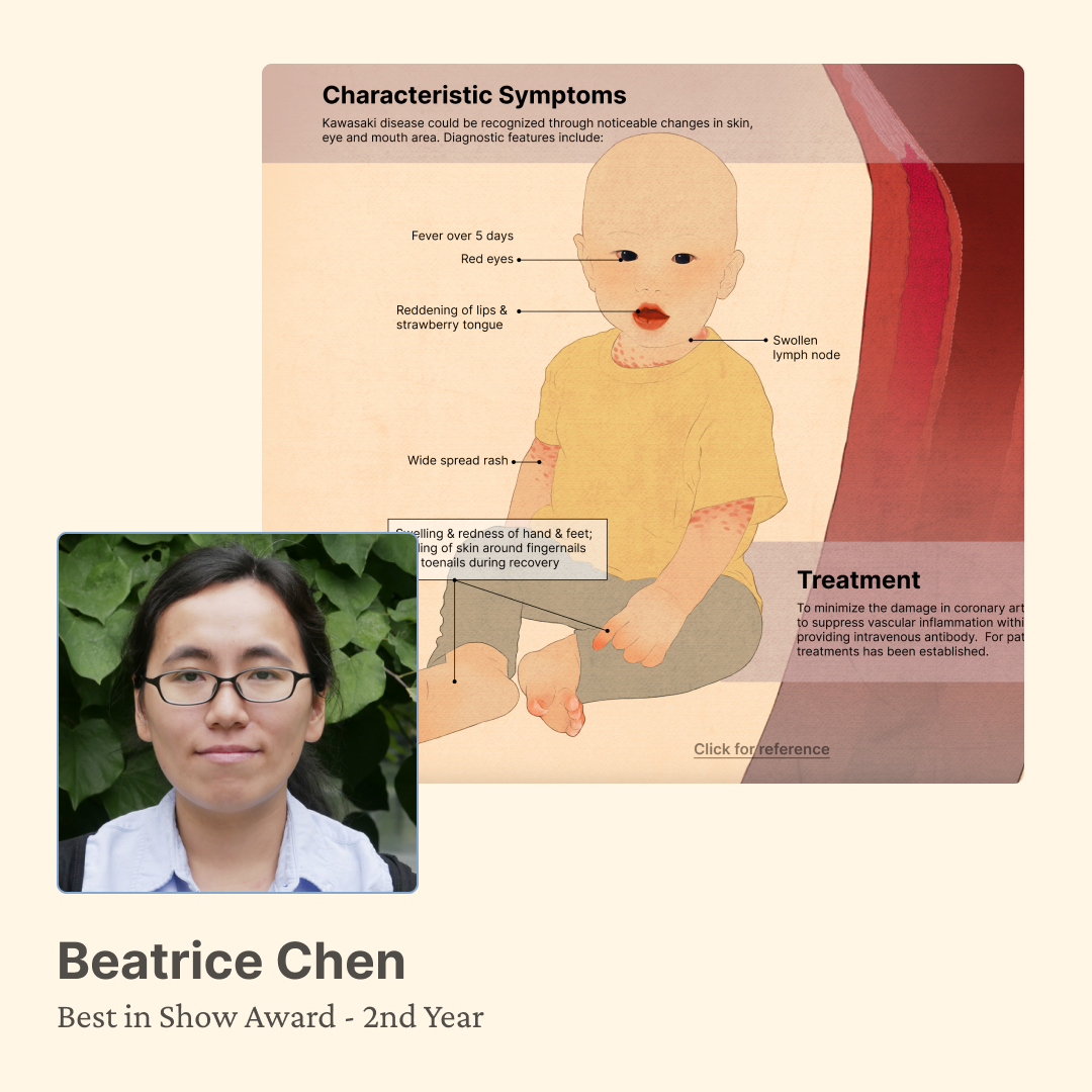
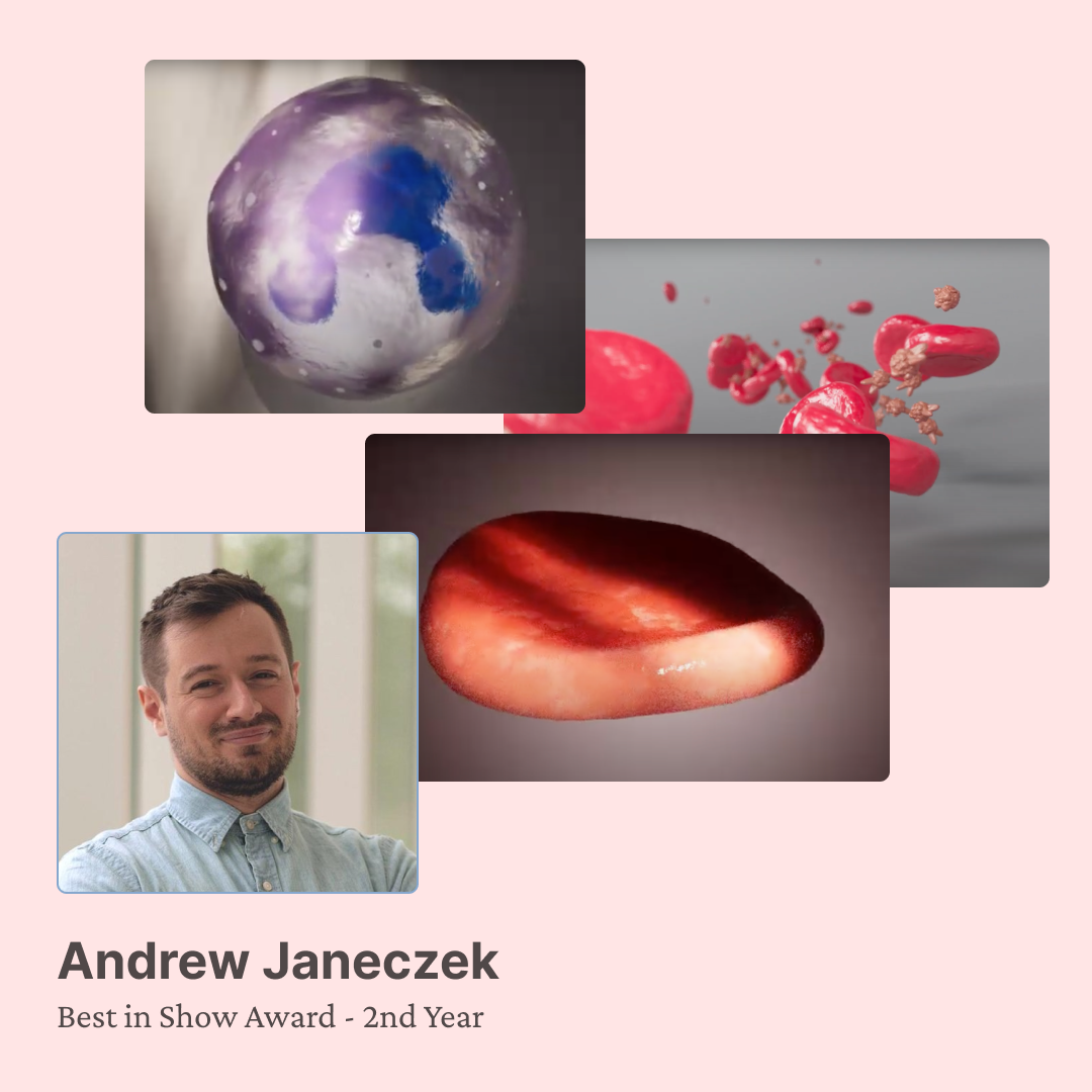
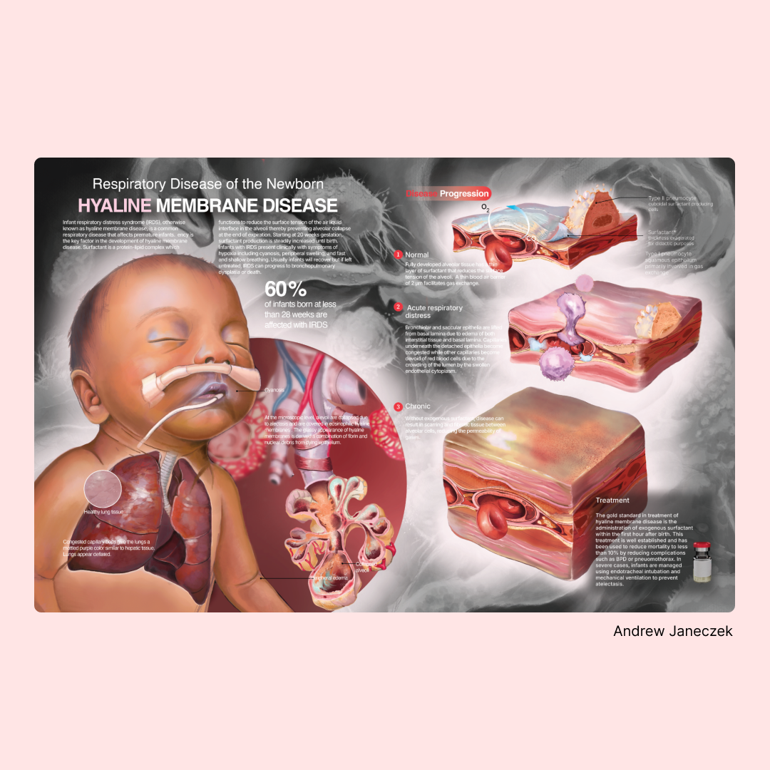
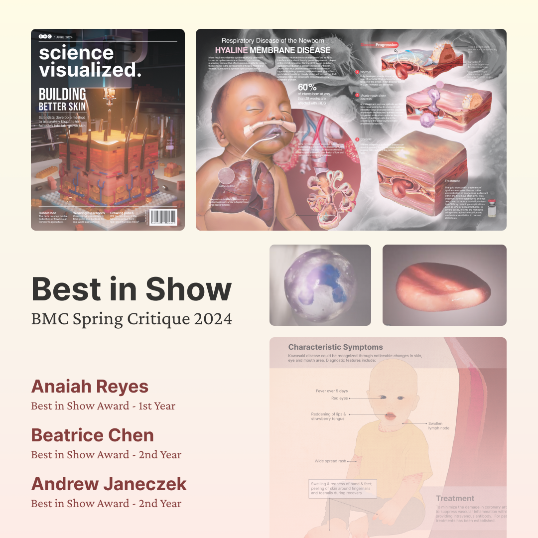
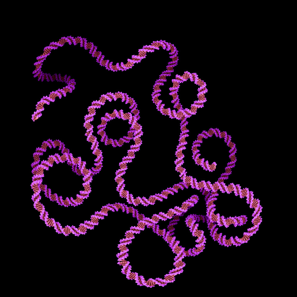
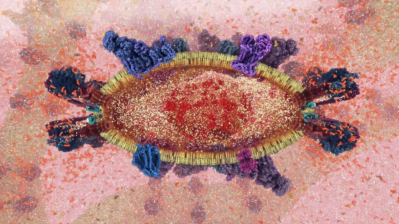
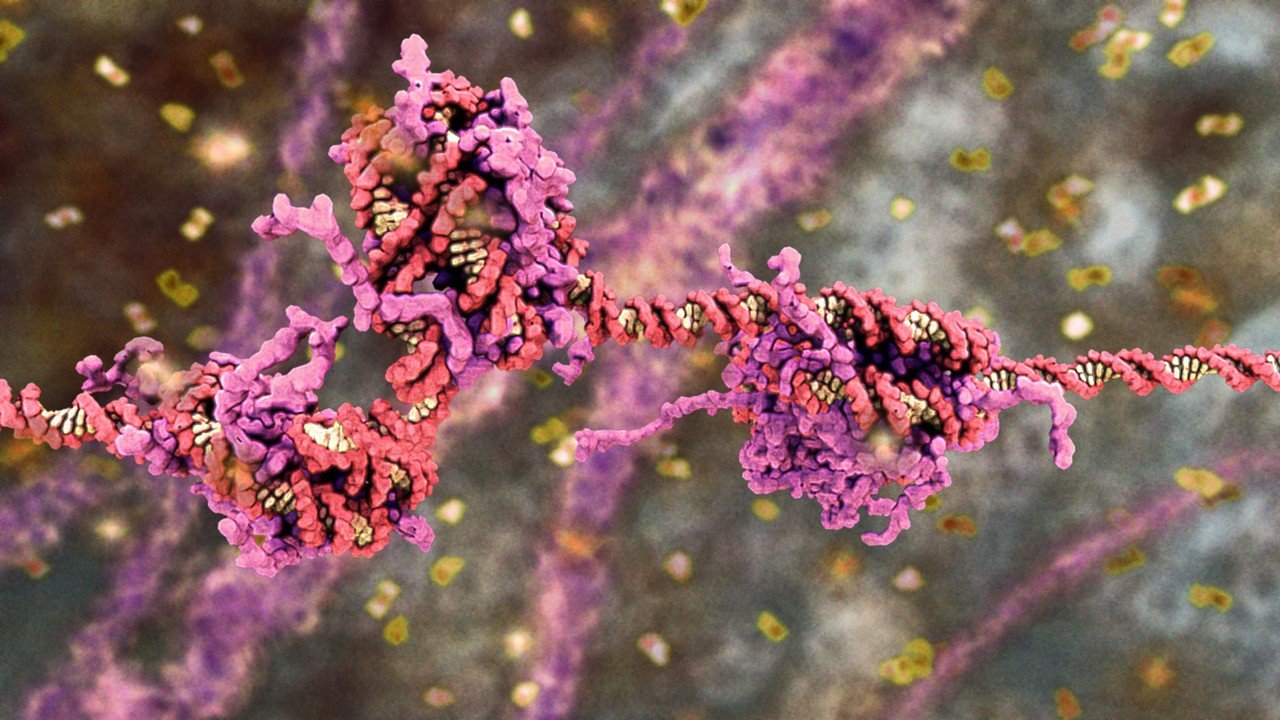
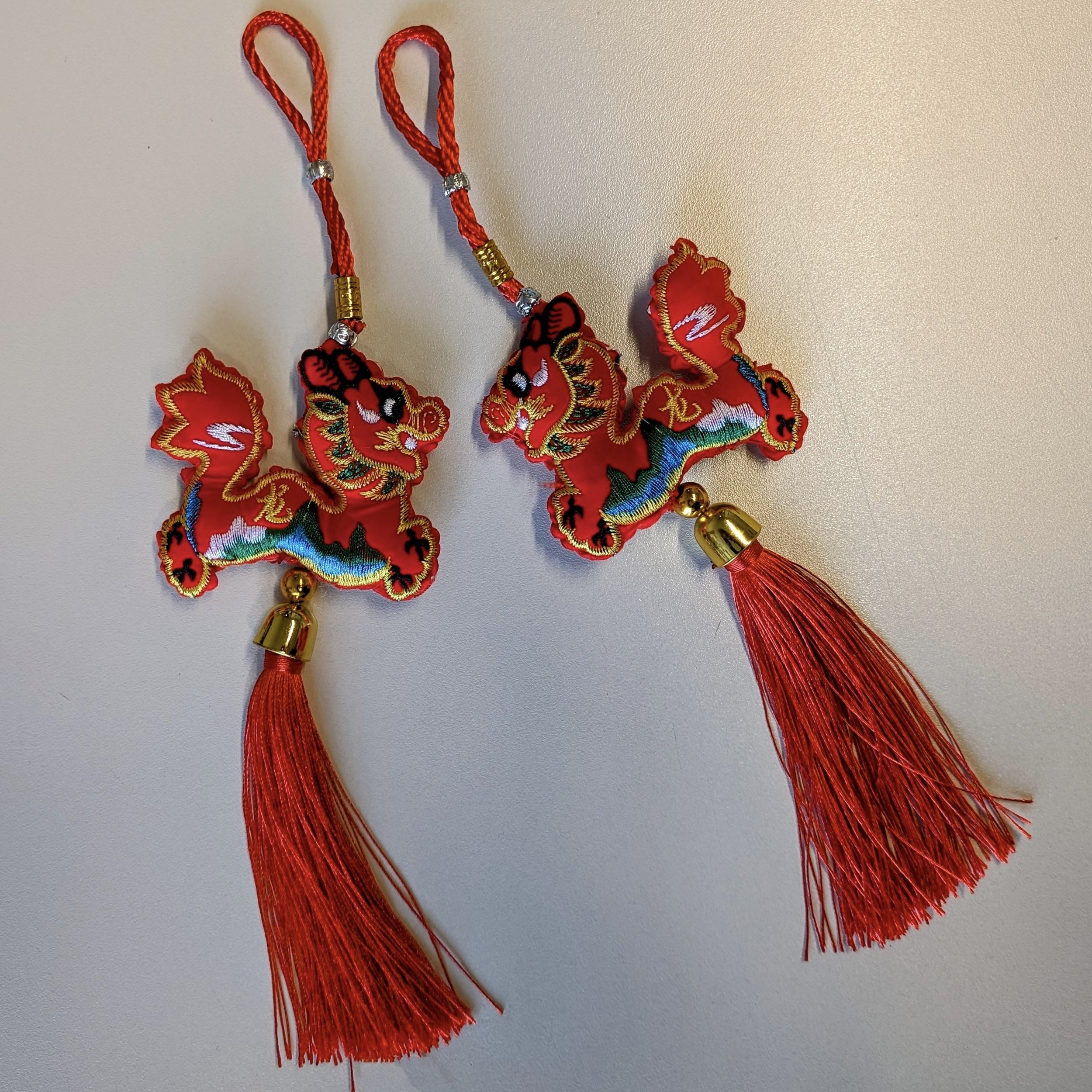
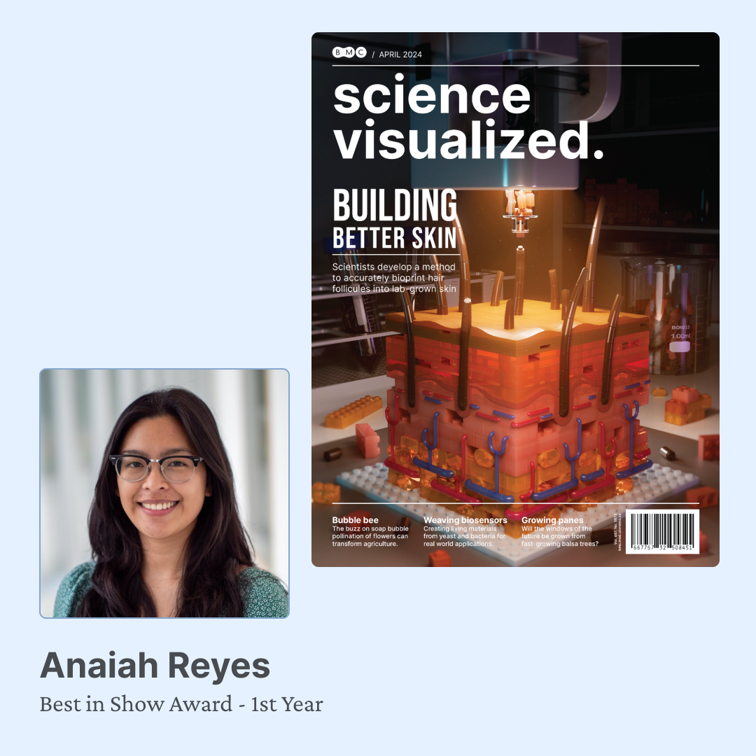
Collage of visualizations from Spring Critique 2024 Best in Show awardees Anaiah Reyes, MScBMC ‘25, Beatrice Chen, MScBMC ‘24 and Andrew Janeczek, MScBMC ‘24.

This past Wednesday, April 17, the first and second year students, biomedical communications faculty, and guest judges, gathered to review student work completed over the winter semester.

Spring Critique is a professional development opportunity where students give and receive constructive feedback on their work–valuable preparation for interacting with clients in industry.

After a morning of reviewing student showcases, attendees voted to name Year I and Year II student showcases the Best in Show. As chosen by their colleagues, Year I's Best in Show was awarded to Anaiah Reyes, MScBMC '25. Beatrice Chen and Andrew Janeczek, both MScBMC '24, tied for Year II's Best in Show.

Thanks to our guest judges Brittany Cheung, MScBMC '21, Professor Emerita Margot Mackay, BScAAM '68, and Michie Wu, MScBMC '22.

Congratulations to our winners and to all our students on the successful completion of the winter semester!

~

Web sites referenced

Beatrice Chen’s online portfolio http://beatricenjc.weebly.com

Andrew Janeczek’s online portfolio https://www.andrewjaneczek.com

Anaiah Reyes’ Instagram https://www.instagram.com/areyes_visuals/

2024 BMC speaker series: Drew Berry, MSc, PhD (hc), Biomedical Animator wehi.tv

Drew Berry, MSc, PhD (hc), Biomedical Animator wehi.tv

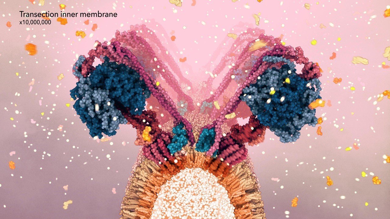
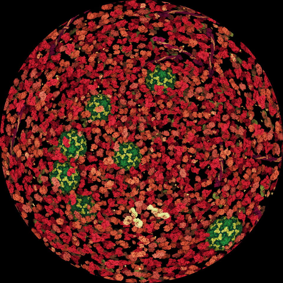
Generative, dynamic model of a Lysosome organelle

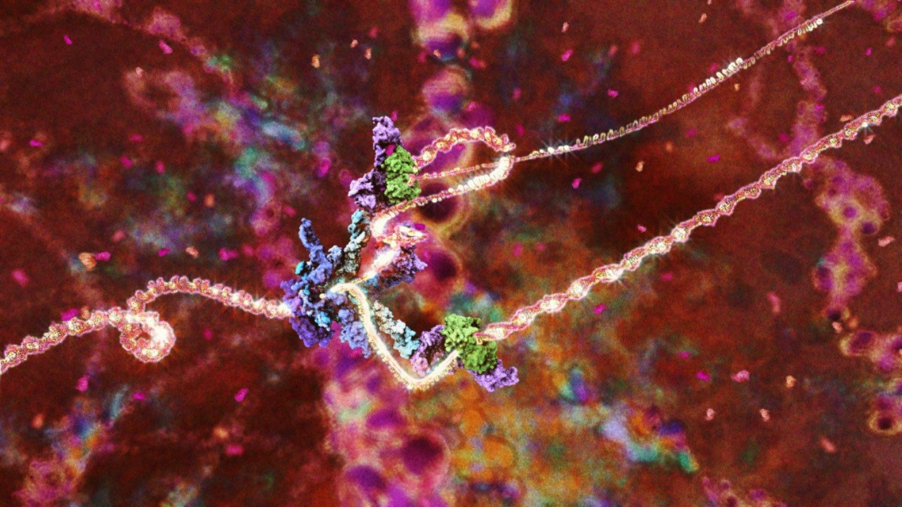
Drew Berry will present wehi.tv’s latest production to generate dynamic, real-time organelle-scale models for science communication and education.

Date: Friday, March 22, 2024
Time: 2:00 p.m. to 3:00 p.m. ET
Register to attend webinar: https://utoronto.zoom.us/webinar/register/WN_FXa6j9vYSeG9QoADJCHmJQ
2024 BMC speaker series program. https://bmc.med.utoronto.ca/news-events/2024/02/01/bmc-speakers-2024


Drew Berry is a biologist-animator renowned for his visually stunning and scientifically accurate animations of molecular and cellular processes. With a background in cell biology and microscopy, Drew ensures that each project is scientifically rigorous and based on current research data. He has been a biomedical animator at WEHI Australia since 1995, and his work has been showcased at international venues such as the Guggenheim Museum, MoMA, and the Royal Institute of Great Britain. In 2011, he collaborated with Björk on her album Biophilia. Drew has received numerous awards, including an Emmy, a BAFTA, and the MacArthur Fellowship Genius Grant.

Web site wehi.tv
YouTube https://www.youtube.com/playlist?list=PLD0444BD542B4D7D9
TED http://www.ted.com/talks/drew_berry_animations_of_unseeable_biology
Architectural projection https://www.youtube.com/watch?v=m9AA5x-qhm8
Björk video https://youtu.be/Yn8AC8z2adU
Wikipedia https://en.wikipedia.org/wiki/Drew_Berry

BMC graduate students help create inclusive new medical curriculum

Standing L-R, MScBMC Class of ‘25 graduate students Yu-wen Jan, Bonnie Wang and Ashton Goebel show their palettes of professional body paints. The medical illustrators painted the lumbar anatomy onto the backs of the nine patient models seated in front of them. Photo credit: Danielle Dilkes for the Health Education Media Library, Western University.

On a recent Saturday in February, three Biomedical Communications graduate students traveled to Western University in London, Ont., to paint the internal anatomy of the lower back onto nine patient models.

Photos and videos of the painted models will be used in the creation of new educational materials that represent a diverse patient population. The inclusive curriculum will be used in training medical residents to perform lumbar punctures.

Courtney Casserly, an assistant professor of neurology in the Department of Clinical Neurology at Western, is leading the medical curriculum development team.

She contacted the Master of Science in Biomedical Communications at the University of Toronto in December 2023 with an inquiry about anatomical body painting.

“I responded to Dr. Casserly that not only did we have the expertise, but we also had the tools and some experience with this kind of thing,” says Shelley Wall, associate professor in the biomedical communications program.

Écorché is a technique in art where the human body’s underlying anatomy is depicted without the skin. Écorché can be represented in painting or sculpture, but it can also be created on a model’s body.

Wall taught the anatomical illustration component of the BMC program’s first-year human anatomy course in fall 2023.

“The knowledge was fresh, so I approached the top three students who were having the most success with anatomy,” Wall says.

Ashton Goebel, Yu-wen Jan and Bonnie Wang all said yes. “No hesitation,” says Goebel.

L-R, Bonnie Wang, Yu-Wen Jan and Ashton Goebel review an illustration of the lumbar anatomy created by Jan. The students used the illustration as a template for reference to expedite painting the models. Credit: Maeve Doyle

To help the students prepare for the opportunity, Wall organized an écorché workshop. All BMC graduate students were invited to participate.

John Tran, anatomist and assistant professor (teaching stream) in the division of anatomy, department of surgery at U of T, teaches the gross anatomy component of the human anatomy course. He delivered refresher lectures to the students on the lumbar spine, and hand and forearm anatomy.

Tran worked with Goebel, Jan and Wang to show them how to identify landmarks on the model that are relevant to lumbar puncture. The students worked for the first time with professional body paint to visualize the bony structure of the lumbar spine onto the living human model.

Other graduate students painted hand and forearm anatomy onto BMC faculty volunteers.

BMC graduate students practice painting the hand and forearm anatomy onto skin. Photo submitted by Shelley Wall

TEACHING LUMBAR PUNCTURE

Casserly teaches the lumbar puncture or LP procedure to medical students and residents in neurology.

“One of the challenges we faced in teaching the LP was that the educational materials focused on young, able-bodied, white men when, in reality, the patients we’re seeing are all sizes, all ages,” says Casserly. 

L-R, Courtney Casserly and Yu-wen Jan identify bony landmarks on a model’s back. Credit: Maeve Doyle

Casserly and her team deliberately focussed on the inclusion of different body types and ages. They selected models from Western’s standardized patient program who had different skin tones, different amounts of body fat, different degrees of skin elasticity, and with the presence of body art. “We've also looked at spinal curvature to see how that impacts our procedure.” 

Casserly says that by painting the underlying anatomy onto the skin’s surface, “learners will be able to visualize what they are doing and also be able to visualize it on different body types. Visual cues for the learners that might help them to see both the horizontal and vertical landmarks and to really envision what's going on under the skin.” 

She says that that is where the biomedical communications program came in. “We needed people who had training in the scientific medical background, but also people who had those artistic skills to be able to execute that vision in a way that was both beautiful and informative for our learners,” says Casserly.

Examples of the lumbar anatomy illustrations at various stages and on a variety of models. Credits: Maeve Doyle

Over five-and-a-half-hours in a Western University nursing simulation suite, Goebel, Jan and Wang painted the lumbar anatomy onto three models each. They painted from the lower rib cage and T12 vertebra down to the tailbone, and the pelvic bones and iliac crests.

Wang says that they used their training from Tran to identify and paint landmarks onto the seated models’ backs as they leaned forward. After landmarking, the medical illustrators created outlines and painted them in.

As prepared as the medical illustrators were, they still experienced some challenges.

“Not every standardized patient could be landmarked the same way because of individual variation in body size and muscle mass in their back. In those cases, I did the best I could and relied on the experts there to make informed approximations,” says Wang.

Bonnie Wang, MScBMC ‘25. Credit: Maeve Doyle

One of the models was a former gymnast. Jan says that “her back muscles were so strong that it was difficult to identify the bony landmark from her muscle. Also, because she was short-waisted, the landmarks were in a different location from where we expected at first.”

Jan, Casserly and anatomist Charys Martin, an assistant professor in Western’s medical school, worked for several minutes to identify the landmarks. Casserly said that this was representative of the real-world experience.

After spending much of the last semester creating only digital visualizations, Goebel says it was good to return to a traditional medium and have a new kind of experience. “Painting on skin is completely different from painting on canvas or paper,” they say.

L-R, Charys Martin and Ashton Goebel, MScBMC ‘25. Credit: Maeve Doyle

“We also had to take into consideration the patient’s comfort. And unlike digital art, there’s no undo button,” says Wang.

REPRESENTATION IN MEDICAL CURRICULUM

“Learners are going to encounter all different patients. They need to know how to approach a person who is young or old. How skin elasticity will impact things. How different spines, curvatures, spaces, levels of degenerative change and even tattoos impact things. If we focus on one specific body type when we teach these medical students, then they’re not ready to approach a lumbar puncture in clinical practice,” says Casserly.

Goebel says that medical illustrators have a responsibility to create materials for not only medical professionals, but patients too, so that they see themselves represented in medical media.

Wall agrees. “Medical illustrators and medical educators are important partners in developing learning materials that represent the broad range of patients that new doctors will meet in their practice.”

The newly created educational tools will be available by the end of summer 2024 through Western University’s Health Education Media Library. The resource is available to anyone around the world who has access to a computer and the internet.

~

Web sites referenced.

Ashton Goebel’s Instagram https://www.instagram.com/ashton.arts.sometimes/

Bonnie Wang’s Instagram https://www.instagram.com/visualsbywang/

Charys Martin’s profile https://www.schulich.uwo.ca/anatomy//people/faculty/faculty_members/martin_charys.html

Courtney Casserly’s profile https://www.schulich.uwo.ca/cns//people/faculty/neurologists/bio-Casserly.html

Health Education Media Library, Western University https://ir.lib.uwo.ca/healthedu/

John Tran’s profile https://surgery.utoronto.ca/faculty/john-tran

Living anatomy workshop 2015 https://bmc.med.utoronto.ca/news-events/2015/04/20/living-anatomy-workshop?rq=living%20anatomy

Shelley Wall’s profile https://bmc.med.utoronto.ca/faculty-staff/#wall

Yu-wen Jan’s Instagram https://www.instagram.com/dinosaur.visuals/
2024 BMC speaker series: Albert Fung, Team Lead, Senior Biomedical Communication Specialist, TVASurg

Albert Fung, MScBMC ‘10

Joining worlds through storytelling

Storytelling has long been an effective communication tool in human history, allowing us to encapsulate information in an engaging manner. As biomedical communication specialists, our unique role allows us to understand the original message, but also to enhance the narrative using various tools in design, aesthetics, and technology. In this talk, I will share some examples of stories we help to tell, from detangling the steps of complex surgeries to empowering patients' understanding of their anatomy, with some tips and tricks along the way.

Date: Friday, February 9, 2024
Time: 2:00 p.m. to 3:00 p.m. ET
Register to attend webinar: https://utoronto.zoom.us/webinar/register/WN_ms4gyuvTRQaMQgTfHnmdDg
2024 BMC speaker series program. https://bmc.med.utoronto.ca/news-events/2024/02/01/bmc-speakers-2024


Albert Fung, MScBMC ‘10, works at the Toronto Video Atlas of Surgery (TVASurg), a small biomedical communications team embedded in the Department of General Surgery at Toronto General Hospital. The team has been producing teaching videos that blend together high-definition footage obtained in the surgical field with patient-specific 3D computer animations. Working with collaborators around the world, the team strives to build a library of surgical cases that span different surgical specialties and that is an open-access resource for students and trainees.

Web site http://tvasurg.ca

SciViz communications skills workshop for biology graduate students: Improve your scientific figures with vector illustration software

The UTM Biology Graduate Student Society is holding a visual communications skills workshop.

Tuesday, February 20, 2024
1 to 4 p.m. ET
Room 3160 CCT Building, UTM

Make better charts, diagrams, cladograms and posters using Adobe Illustrator and/or Affinity Designer.

Space limited so REGISTER to attend! https://forms.office.com/r/TzB35tZJuP

~

Your host Dave Mazierski is an expert in image creation, vector graphics and medical illustration. He is the creator of hundreds of medical and scientific illustrations for books, posters and education.

A faculty member in the Master of Science in Biomedical Communications program, Dave teaches human anatomy, traditional medical illustration techniques, 2D digital illustration with Adobe Illustrator and Photoshop, applied art in pathology, and the history of medical illustration.

2024 Stephen Gilbert Award winners

MScBMC Class of ‘24 graduate students Natalie Lucas (left) and Juno Xi Shemano (right) received this year’s Stephen Gilbert Award for Artistic Excellence.

On Tuesday, February 6, 2024, the BMC program announced the winners of the Professor Stephen Gilbert Award for Artistic Excellence in Biomedical Visualization.

Natalie Lucas and Juno Shemano won for their collaborative work Head & Neck: Cranial Nerve V. Lucas created the linework and layout for the winning piece, and Shemano rendered the final image. Lucas and Shemano are both second-year students in the BMC program.

Head & Neck: Cranial Nerve V–Stephen Gilbert Award-winning piece created by Natalie Lucas and Juno Xi Shemano, both MScBMC Class of ‘24.

The Stephen Gilbert Award is made annually to a BMC graduate student who demonstrates artistic excellence in the representation of structure and function at the gross, micro or molecular level. Work submitted for consideration for the award must have been completed within a BMC course.

The award announcement was made at the BMC program’s 2024 Lunar New Year social.

Congratulations Natalie Lucas and Juno Shemano on your award of merit!

~

Websites referenced

MScBMC Awards https://bmc.med.utoronto.ca/mscbmc-awards

Natalie Lucas’ portfolio https://www.natalielucasyoungblood.com

Juno Shemano’s portfolio https://www.junoshemano.com

2024 Lunar New Year social https://bmc.med.utoronto.ca/news-events/2024/2/6/2024-lny-social

2024 Lunar New Year social

Year of the Dragon 2024 .gif created by Yu-wen Jan, MScBMC Class of ‘25

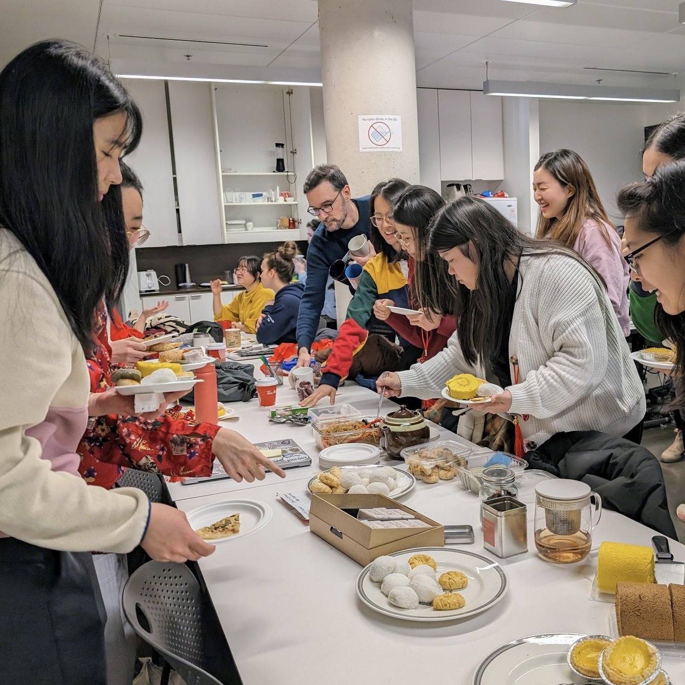
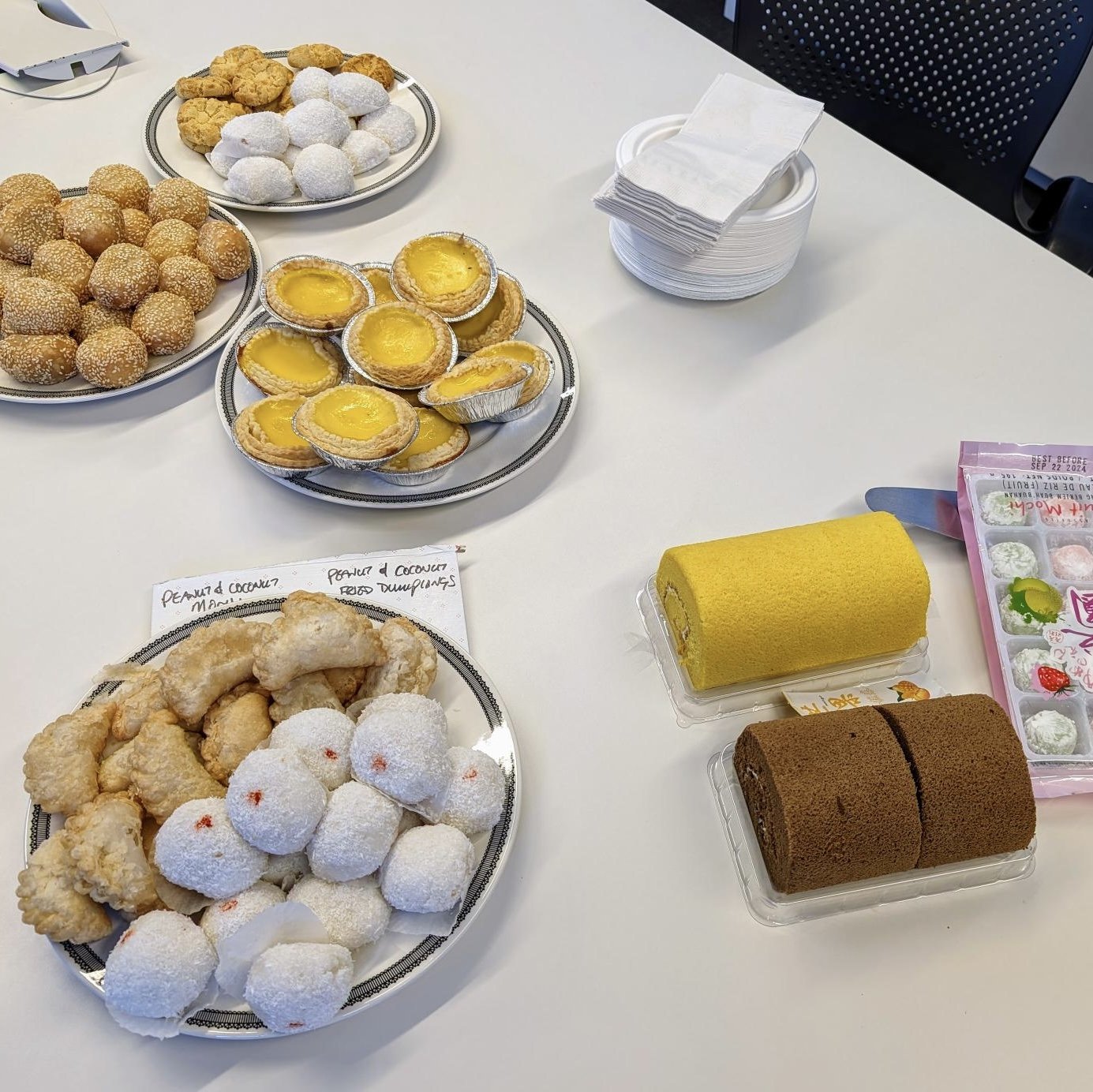
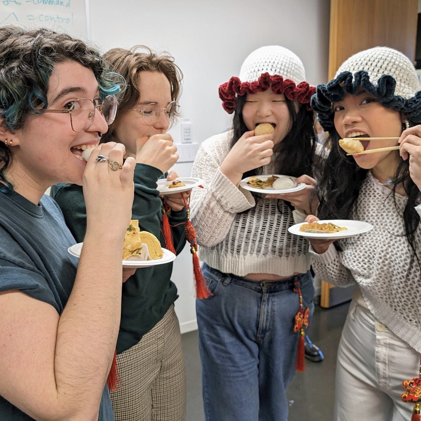
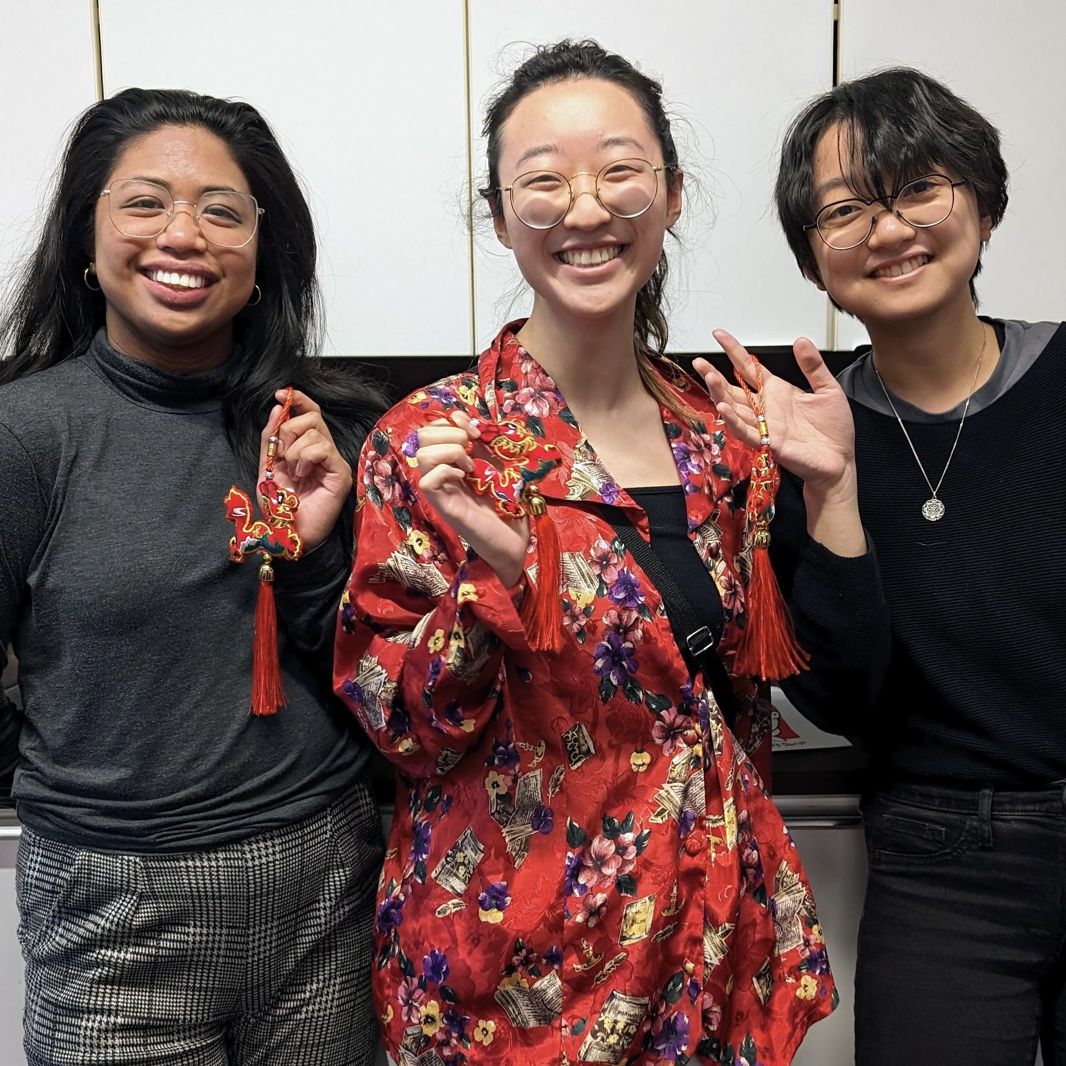
On Tuesday, February 6, 2024, the BMC program welcomed the Year of the Dragon a few days early with tea, traditional pastries, and colourful red-and-gold dragons.

At the social, the winners of the Professor Stephen Gilbert Award for Artistic Excellence in Biomedical Visualization were announced. Congratulations second-year students Natalie Lucas and Juno Shemano.

Special thanks to BMC graduate student Yu-wen Jan for our bespoke Year of the Dragon 2024 .gif, Professor Dave Mazierski and Year 2 Student Rep Samantha Li for the pastries, and Professor Shay Saharan for the tea.

Happy Lunar New Year!

~

Websites referenced

MScBMC Awards https://bmc.med.utoronto.ca/mscbmc-awards

2024 Stephen Gilbert Award winners https://bmc.med.utoronto.ca/news-events/2024/2/6/2024-stephen-gilbert-award-winners

Yu-wen Jan’s Instagram https://www.instagram.com/dinosaur.visuals/

BMC Faculty & Staff https://bmc.med.utoronto.ca/faculty-staff

Samantha Li’s portfolio https://www.samlivisuals.com


2023 Wendy M. Kates Award recipients

Members of the Kates family attended the Class of 2023’s Final Presentations on November 7, 2023. Jerry and Shawn Kates personally congratulated Livia Nguyen and Linda Ding for receiving the 2023 Wendy M. Kates Memorial Award for their master’s research project. Left to right: Wendy Kates’ father Jerry Kates, Livia Nguyen, Linda Ding, Wendy Kates’ brother Shawn Kates, MScBMC program director Jodie Jenkinson. (Photo courtesy of Jerry Kates.)

Congratulations to Nan-Linda Ding and Livia Nguyen the 2023 co-recipients of the Wendy M. Kates Memorial Award. The Master of Science in Biomedical Communications program makes this award on the basis of academic achievement in the master's research project and commitment to the profession.

Ding and Nguyen, MScBMC '23, were recognized for their design and creation of the 2D narrative educational video game NavEDI. NavEDI teaches clinical advocacy, and educates medical students about equity, diversity and inclusion in the psychiatric ER.

The Biomedical Communications program is grateful to the Kates family who established this award in Wendy’s memory and who have worked tirelessly for decades to provide the principal funding for the award.

Donations to the Wendy M. Kates Memorial Award can be made here. https://engage.utoronto.ca/site/SPageServer?pagename=donate#/fund/915

MScBMC Open House 2023

Date & Location

Saturday, September 16, 2023 from 10 a.m. to 1 p.m.

University of Toronto Mississauga (Details to be provided when registrations are confirmed.)

For directions and campus maps, see https://bmc.med.utoronto.ca/visit-us.

Drop-in

The BMC Open House is an informal, casual opportunity for potential applicants to meet with students who are currently enrolled in the program, to tour BMC's facilities at UTM, and to meet with faculty. Potential applicants can request a pre-application review to help them assemble the strongest application possible when they do apply. This is not an interview. Attendance at the Open House is not a guarantee of admission.

Formal interviews for Fall 2024 admission will take place February 22, 2024 and are by invitation only. (See Important Dates.) Ideally, interviews will be in person. However, if interviews must take place on-line, the BMC Open House will have provided an opportunity for potential applicants to meet BMC faculty in person and to seek feedback on transcripts and art.

Pre-application review

For potential applicants who would like a pre-application review, there are available a limited number of 10-minute scheduled appointments with various faculty members.

Review the BMC faculty profiles and request an appointment with someone whose areas of specialization are of interest to you. https://bmc.med.utoronto.ca/faculty-staff

Required

Attendees must register to attend, and must bring unofficial transcripts and art samples with them. Register here: https://forms.office.com/r/wGQcGkF1xr