
Brendan (1T4) Polley develops “virtual hands” to manipulate digital 3D anatomical models.
Illustrating Medicine: An Exhibition of Original Medical Illustrations from J.C.B. Grant’s Atlas of Anatomy

Illustrating Medicine: Art exhibit that features original artworks from the MScBMC archives
Vernissage: Thursday, March 13, 2014 from 4:30 to 7:30 pm
Exhibit: March 13 to May 1, 2014
Location: CJ Building, Loyola Campus, Concordia University, 7141 Sherbrooke Street West, Montreal, Media Gallery, Room 1.419
Stephen Goltra Gilbert 1931-2014
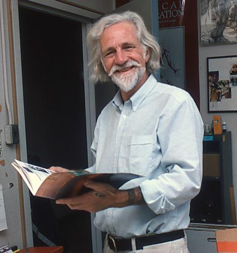
Photo of Steve Gilbert in 1994
With great sadness, we announce the passing of Stephen Goltra Gilbert.
Steve was born in Portland, Oregon on January 18, 1931. As a child, he was mesmerized by animated cartoons, and his parents encouraged his artistic nature and his love of the natural world.
While at boarding school in Andover, Massachusetts, Steve discovered musical theatre and opera. He then attended Reed College where he earned a degree in art, and was introduced by a colleague to the work of Frank Netter, which fascinated him. During a three-year stint in the U.S. Army Medical Corps, he applied to study medical illustration under Muriel McLatchie Miller (a former student of Max Brödel) at Massachusetts General Hospital, which he started following his military discharge. After three years of executing drawing after drawing of human anatomy, Steve received his first commission from a surgeon in Tacoma, Washington, who arranged for Steve to receive additional training from Ralph Sweet in San Francisco. Steve was grateful for the two months he spent with Sweet and learned a great deal, but his frustration with institutional illustration work led him to leave employment with the University of Washington’s School of Medicine and return to the family farm in Oregon, where he spent the next twelve years writing and illustrating his greatest body of work: six dissection guides for biology (necturus, frog and atlas of comparative zoology; also fetal pig, cat and dogfish, which are still in print) based on his own research and dissections.
In 1973, Steve was invited to Toronto, where he was offered a position to teach part-time applied art courses in the Art as Applied to Medicine Department in the Faculty of Medicine. Over his twenty-three year career at U of T, he attained the title of full professor and instructed over one hundred AAM and BMC students. He also authored “Pictorial Human Embryology” (1989) and “Outline of Cat Anatomy With Reference to the Human” (1999). He was the recipient of many AMI and teaching awards, and was invited to Japan on multiple occasions to train young illustrators there. Steve never completely retired; among other projects, he continued to work on an anatomy book with Dr. Anne Agur at the University of Toronto and completed a pictorial history of embryology, which is still awaiting a publisher. He will also be remembered as a tattoo artist and historian, who authored many articles and a book on the history of tattoos. Many of his friends, former students and family members carry their own indelible, permanent reminder of Steve’s legacy with them. His beautiful tone and pen & ink illustrations, his gentle and caring nature, and his great passion for art, science and truth will always inspire us to be better illustrators, teachers, and human beings.
There will never be another man like him. Steve died in his sleep in Toronto on February 21, 2014 after a long illness. He is mourned by his wife Cheralea and their children Scott, Genevieve and Emily, and his children Tom, Ann, and David.
by Dave Mazierski
Educational Design of eLearning Environments for Health Care Professionals

Presentation by MScBMC Faculty, Dr. Leila Lax
“Educational Design of eLearning Environments for Health Care Professionals”
Wednesday, March 5, 2014 from 12 noon to 1 pm
Women’s College Hospital – Level 6, Room 6214
76 Grenville Street, Toronto, Ontario M5S 1B1
Insect Drawing Workshop

leptoglossus occidentalis
You’re invited to an insect drawing workshop with MScBMC alumna Kathryn Chorney (9T8).
Join this science/nature/medical illustrator at the Nook Collective for an afternoon of insect drawing.
Sunday, November 17, 2013
12:30 to 4:30 pm
Nook, 158 Augusta Avenue, Toronto, ON M5T 2L4
$55
BMC Unplugs

Participate in ‘Unplugged Hour’. To coincide with World Mental Health Day, UTM’s Health & Counselling Centre encourages everyone to unplug.
Unplugged Hour
Thursday, October 10, 2013
12 noon to 1 pm
Disconnect from your phones, computers and social media.
Get outside, sketch, spend quality time with those around you.
Learn more:
https://www.facebook.com/UnpluggedHour
Here’s to good mental health. :)
BMC Alumni Association UnCon 2013

The BMCAA ‘Unconference’ is back for its seventh instalment.
Join your peers on Saturday, November 9 from 10 AM to 3 PM for a lively UnCon. This is a participant-driven event where you, the attendees, share ideas about biomedical visualization.
Have something interesting to share? New adventures with new media? New techniques? If you think any of your fellow BMCAA members would be interested, send us your presentation idea!
Don’t feel like presenting? No problem! Come down and enjoy a day of networking and learning.
During lunch, alumni are welcome to convene for the BMCAA Annual General Meeting. The meeting will be a forum for the discussion of alumni association issues, including the election of a new BMCAA newsletter editor.
A website with additional information, including login access to sign up as an attendee or a presenter will follow shortly. We look forward to seeing you all at the UnCon this year!
BMC Alumni Association Executive Committee
bmcaa.exec@gmail.com
Visualizing the invisible

“The inner ear is one of the toughest topics to teach because it is anatomically very complex, very small and buried in the dense petrous bone,” said Professor Emerita Pat Stewart.
Andrea Zariwny, a Biomedical Communications graduate student, tackled this teaching problem with an augmented reality iPad app and 3D-printing to visualize the anatomy of the inner ear.
Here’s how it works: Launch Zariwny’s app. Hold the iPad over Zariwny’s illustration of the petrous bone. The illustration acts as a glyph—a high-contrast image recognized by the tablet’s camera—and triggers a 3-dimensional digital model on top of the illustration. Look through the digital model to the illustration below and see the cochlea of the inner ear or the spiral ganglion lift right off the page.
“The illustration on its own is an informative, didactic tool,” said Professor Marc Dryer, Zariwny’s research supervisor. “But augmented with another layer of information, it becomes a strong educational feature that enhances the learning experience and engages student attention.”
Zariwny and her content advisor, Stewart, used micro CT scanning to create medical images from real anatomical evidence. Zariwny took the data into a medical image viewer and extracted 3D digital models of the petrous bone, and the cochlea and spiral ganglion—internal structures of the inner ear. She retopologized or ‘cleaned up’ the models and took them into gaming software to create the 3-dimensional digital models.
After Zariwny, who has a background in industrial design, created the augmented textbook illustration, she wanted to work with 3D printing. “I thought augmented reality could be an even more useful tool combined with a physical model,” said Zariwny.
Zariwny 3D-printed a model of the petrous bone. The model rests on a separate platform—a plinth. She wrapped the plinth in a glyph. She printed another petrous bone but, this time, sliced in half and printed the glyph onto the cross-sections. She created modules for her app that “ghost” the internal structure of the inner ear over the physical models. The apps recognize and track the glyphs, and match the digital models to the position of the physical models. Move the models, or the iPad, and the digital models allow viewers access to different orientations and perspectives.
Zariwny’s tool accurately conveys the size and position of what is essentially space buried in bone, said Stewart. “The first time I saw the models, I thought, that’s it! Exactly what we need.”
by Maeve Doyle
Pratt Manhattan Gallery to host Splice exhibit

Pratt Manhattan Gallery hosts “Splice: At the intersection of Art and Medicine” curated by Nina Czegledy.
September 20 to November 9, 2013
PRATT MANHATTAN GALLERY
144 West 14th St. | New York, NY 10011
212.647.7778 | exhibits@pratt.edu | www.pratt.edu/exhibitions
Opening Reception: Thursday, September 19, 6 to 8 pm. Special event, opening night only: Explore Asian health traditions and immortality with wild mushroom hunter Med Shroomdog at the Mushroom Tea Bar.
Showcasing 20th-century anatomical drawings complemented by contemporary works of art, SPLICE: At the Intersection of Art and Medicine brings together both scientific and artistic means of representing the human body. It also represents the first significant public exhibition within the United States of anatomical images selected from an extensive collection housed at Biomedical Communications, University of Toronto. The University of Toronto Art Center, the Blackwood Gallery, University of Toronto, Mississauga, and the West Vancouver Museum presented previous versions of this exhibition.
Anatomical Artists: Elizabeth Blackstock, Dorothy Foster Chubb, Marguerite Drummond, Nancy Joy, Eila Hopper-Ross, Maria Wishart
Contemporary Artists: Ælab: Gisèle Trudel and Stéphane Claude, Jack Burman, Jack Butler, Andrew Carnie, Joyce Cutler-Shaw, Dana Claxton, Orshi Drozdik, Eric Fong, Terry Kurgan, Patricia Olynyk, Piotr Wyrzykowski
Pratt Manhattan Gallery is a public art gallery affiliated with Pratt Institute featuring exhibitions of art, architecture, fashion, and design.
Nancy Grahame Joy (1920-2013)
Medical artists should be “born teachers, artists by vocation and scientists by nature.”
— Nancy Joy, 1974
Nancy Jean Hannah Grahame Joy, Chair of the University of Toronto’s Department of Art as Applied to Medicine (AAM) (now Biomedical Communications) from 1962 to 1985, passed away on July 27, 2013, in Toronto. As a medical illustrator, she is best known for her contributions to J.C.B. Grant’s Method of Anatomy and Atlas of Anatomy; she worked with Dr. Grant for over thirty years, and was influenced by his views on the importance of illustration as a tool for teaching and research. As a professor, she was an influential leader in shaping medical illustration as an academic discipline. Prof. Joy was one of a few female Chairs of a department at the University of Toronto in the ‘60s. Under her guidance AAM was elevated from a diploma program to a three-year Bachelor of Science in 1967.
Daughter of an artist and a lawyer, and grand-daughter of a surgeon, anatomist, and Dean of the UofT Faculty of Medicine, Dr. Alexander Primrose, Joy combined art and medicine throughout her life. In 1939 she enrolled in a four-year diploma at the Ontario College of Art; she later attended classes in anatomy, histology, embryology, and neuroanatomy with U of T’s medical students, and, from 1944-1946, studied and worked with Tom Jones in the Department of Medical and Dental Illustration at the University of Illinois at Chicago. Upon her return to Toronto in 1946, she enrolled as a special student in the AAM program and worked as a freelance illustrator for Dr. Grant.
Prof. Joy was an accomplished pen and ink and half-tone watercolour practitioner, and she was a forward thinker: as early as 1982 she introduced a course in the design of computer-aided learning for medical education within the AAM program. She was also an active advocate for artists’ rights.
Prof. Joy was an Honorary Fellow of the Ontario College of Art (1984) and a member of the Association of Medical Illustrators. The AMI honoured Nancy Joy and another of Grant’s artists, Dorothy Foster Chubb, at their annual meeting in 1998 in Toronto, when the publisher of Grant’s Atlas presented the original artwork used in the book to Biomedical Communications and the Department of Anatomy. Her legacy as an artist, educator, and advocate continues to flourish in the BMC program and through her medical illustrations in Grant’s Atlas, now in its thirteenth edition.
Donations in honour and in memory of Prof. Nancy Grahame Joy can be made to the Biomedical Communications Program Fund.
Click here to donate online
(Please use BMC address below for online requests for cards; they will be collected by BMC for the family.) To ensure your donation is correctly allocated, please indicate “BMC-Joy” at the end of the online form, under Additional Information.
or
You can donate by cheque. Make cheques payable to the ‘University of Toronto’. To ensure your donation is correctly allocated, please indicate “BMC-Joy” on your cheque and mail to:
Donations
Biomedical Communications
Room 308, Terrence Donnelly Health Sciences Complex
University of Toronto Mississauga
3359 Mississauga Road North
Mississauga, Ontario L5L 1C6
Tax receipts will be issued by the University of Toronto and sent to the address you provide.
Prof. Leila Lax, Prof. Margot Mackay, Prof. Shelley Wall & Maeve Doyle
(BMC Fundraising Committee)