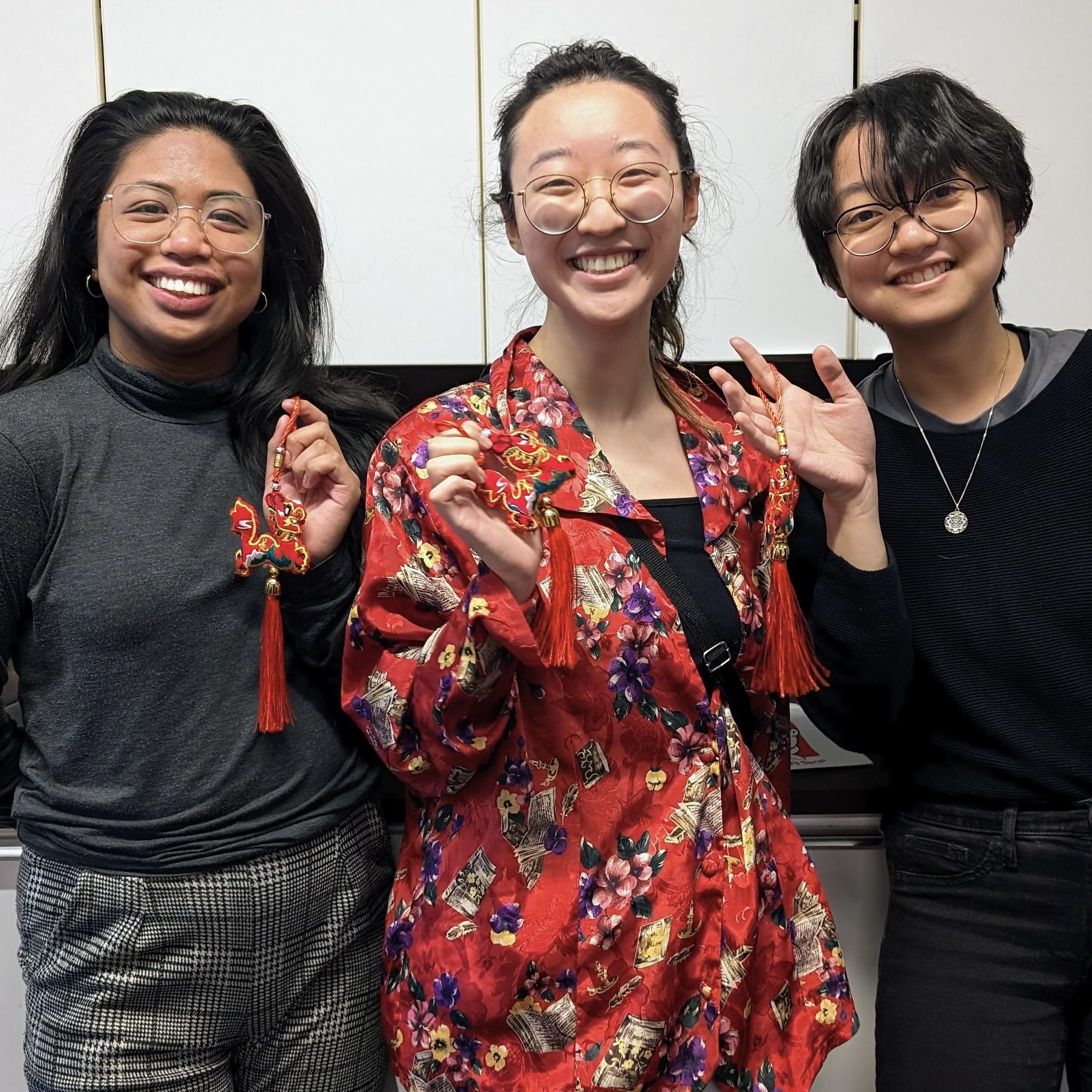
2024 Lunar New Year social

Year of the Dragon 2024 .gif created by Yu-wen Jan, MScBMC Class of ‘25

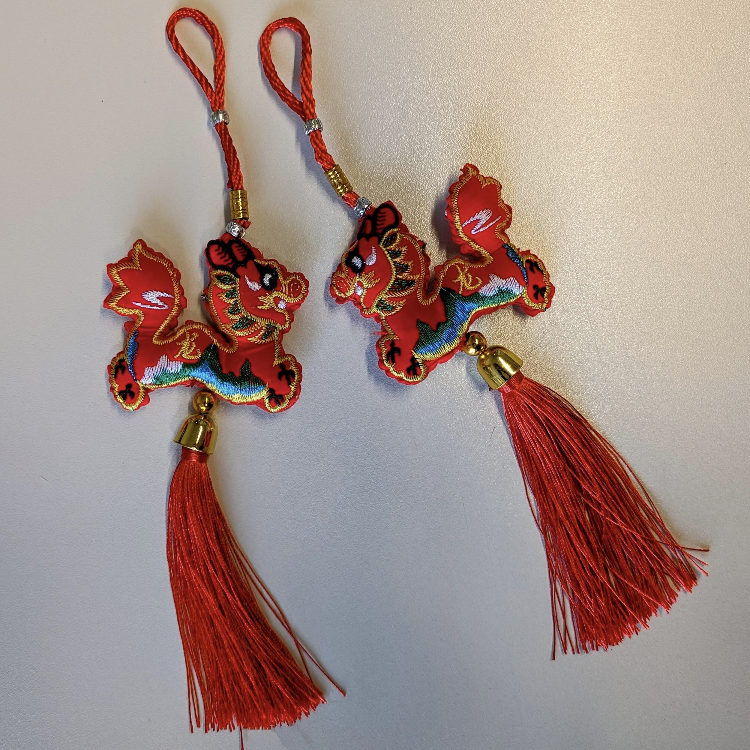
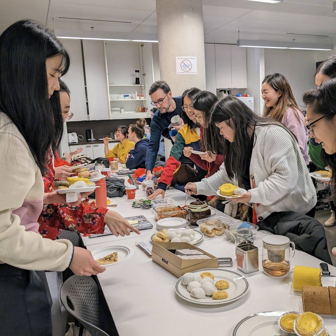
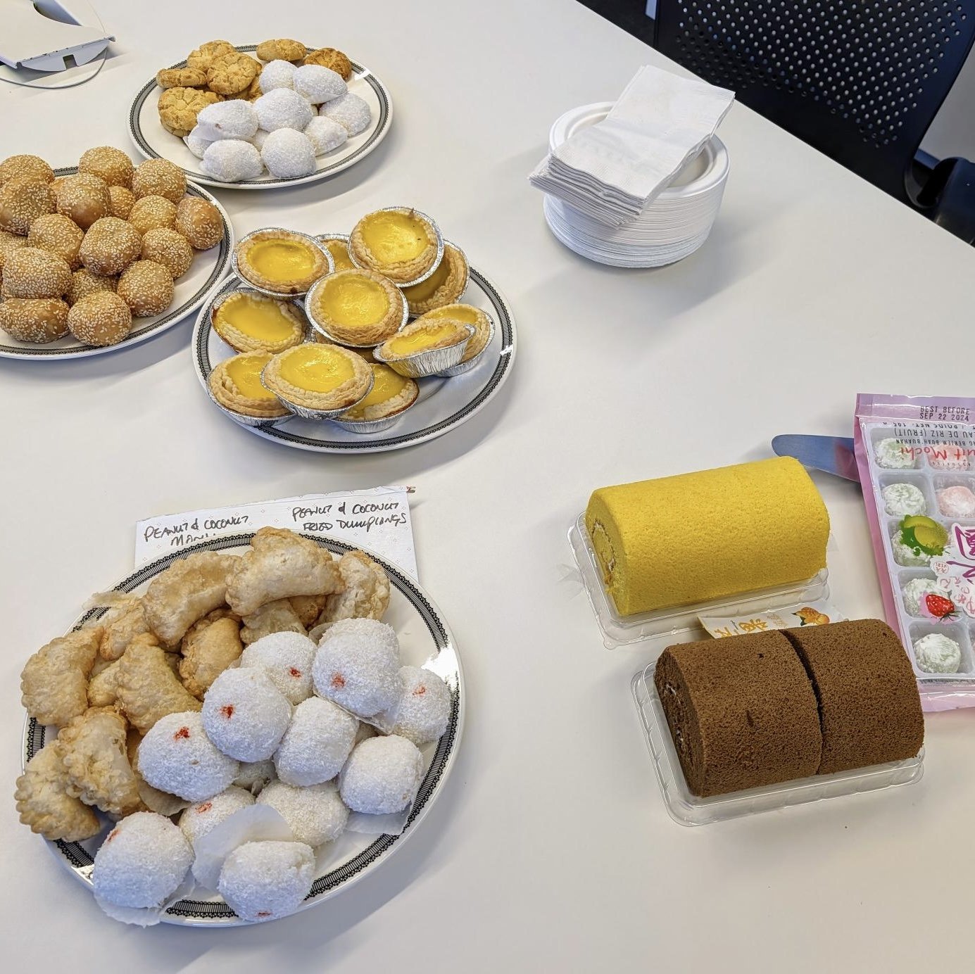
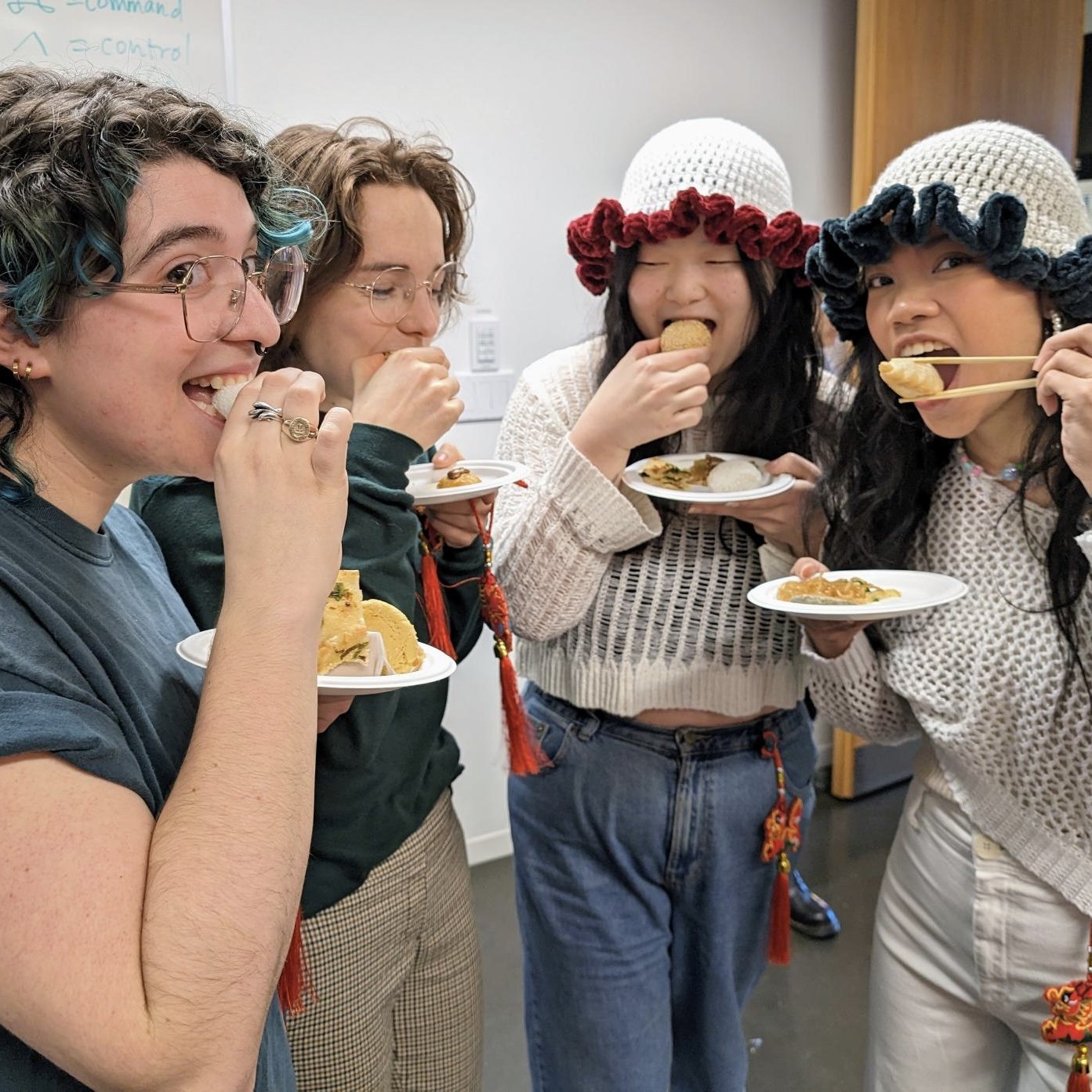
On Tuesday, February 6, 2024, the BMC program welcomed the Year of the Dragon a few days early with tea, traditional pastries, and colourful red-and-gold dragons.

At the social, the winners of the Professor Stephen Gilbert Award for Artistic Excellence in Biomedical Visualization were announced. Congratulations second-year students Natalie Lucas and Juno Shemano.

Special thanks to BMC graduate student Yu-wen Jan for our bespoke Year of the Dragon 2024 .gif, Professor Dave Mazierski and Year 2 Student Rep Samantha Li for the pastries, and Professor Shay Saharan for the tea.

Happy Lunar New Year!

~

Websites referenced

MScBMC Awards https://bmc.med.utoronto.ca/mscbmc-awards

2024 Stephen Gilbert Award winners https://bmc.med.utoronto.ca/news-events/2024/2/6/2024-stephen-gilbert-award-winners

Yu-wen Jan’s Instagram https://www.instagram.com/dinosaur.visuals/

BMC Faculty & Staff https://bmc.med.utoronto.ca/faculty-staff

Samantha Li’s portfolio https://www.samlivisuals.com


2023 Wendy M. Kates Award recipients

Members of the Kates family attended the Class of 2023’s Final Presentations on November 7, 2023. Jerry and Shawn Kates personally congratulated Livia Nguyen and Linda Ding for receiving the 2023 Wendy M. Kates Memorial Award for their master’s research project. Left to right: Wendy Kates’ father Jerry Kates, Livia Nguyen, Linda Ding, Wendy Kates’ brother Shawn Kates, MScBMC program director Jodie Jenkinson. (Photo courtesy of Jerry Kates.)

Congratulations to Nan-Linda Ding and Livia Nguyen the 2023 co-recipients of the Wendy M. Kates Memorial Award. The Master of Science in Biomedical Communications program makes this award on the basis of academic achievement in the master's research project and commitment to the profession.

Ding and Nguyen, MScBMC '23, were recognized for their design and creation of the 2D narrative educational video game NavEDI. NavEDI teaches clinical advocacy, and educates medical students about equity, diversity and inclusion in the psychiatric ER.

The Biomedical Communications program is grateful to the Kates family who established this award in Wendy’s memory and who have worked tirelessly for decades to provide the principal funding for the award.

Donations to the Wendy M. Kates Memorial Award can be made here. https://engage.utoronto.ca/site/SPageServer?pagename=donate#/fund/915

MScBMC Open House 2023

Date & Location

Saturday, September 16, 2023 from 10 a.m. to 1 p.m.

University of Toronto Mississauga (Details to be provided when registrations are confirmed.)

For directions and campus maps, see https://bmc.med.utoronto.ca/visit-us.

Drop-in

The BMC Open House is an informal, casual opportunity for potential applicants to meet with students who are currently enrolled in the program, to tour BMC's facilities at UTM, and to meet with faculty. Potential applicants can request a pre-application review to help them assemble the strongest application possible when they do apply. This is not an interview. Attendance at the Open House is not a guarantee of admission.

Formal interviews for Fall 2024 admission will take place February 22, 2024 and are by invitation only. (See Important Dates.) Ideally, interviews will be in person. However, if interviews must take place on-line, the BMC Open House will have provided an opportunity for potential applicants to meet BMC faculty in person and to seek feedback on transcripts and art.

Pre-application review

For potential applicants who would like a pre-application review, there are available a limited number of 10-minute scheduled appointments with various faculty members.

Review the BMC faculty profiles and request an appointment with someone whose areas of specialization are of interest to you. https://bmc.med.utoronto.ca/faculty-staff

Required

Attendees must register to attend, and must bring unofficial transcripts and art samples with them. Register here: https://forms.office.com/r/wGQcGkF1xr

Interactive web-based learning tool Bone Bio named Best of Show at AMI Salon 2023

Bone Bio - An interactive and animated web-based application for learning human osteology developed by Amy Assabgui, MScBMC ‘22. Still image provided by A. Assabgui

Bone Bio, a web-based interactive learning tool developed by Amy Assabgui, MScBMC ’22, was named the Orville Parkes Best of Show in the Student Category for Traditional Media at the July 2023 Association of Medical Illustrator (AMI) Salon in Henderson, Nevada. Bone Bio was also recognized with an AMI Award of Excellence in the Student Category for Motion and Interactive Media.

A web-based human osteology learning tool, Bone Bio combines 3D models with animation and interactivity to visualize and communicate to undergraduate forensic anthropology students the structural relationships of bone.

"Forensic anthropologists require a solid understanding of bone biology,” says Assabgui. “It is required for performing advanced techniques such as classifying age, identifying pathologies and differentiating human from non-human bone."

Tracy Rogers, director of the Forensic Science program at the University of Toronto Mississauga, and then-instructor of third-year osteology, saw that students were struggling to learn bone biology.

"Students have difficulty understanding how the cells and structures of bone relate to one another, where they are found, the level of magnification needed to see them and how they interact," says Rogers.

Rogers reached out to the Master of Science in Biomedical Communications with this learning challenge, and Assabgui responded.

Amy Assabgui, MScBMC ‘22, is a freelance biomedical illustrator and designer based in Seattle, Washington. Photo provided by A. Assabgui

Assabgui surveyed the forensic anthropology students. Her results showed that students were perplexed by the three-dimensional relationships of bone across different scales, and that they lacked access to real bone material for practical analysis. She found that there were no supplemental learning resources available and no dedicated laboratory component where students could examine bone material.

She developed Bone Bio so students could interact, at their own pace, with 3D models of bony structures, tissue types and multi-layered structures of bone at different levels of magnification. She divided the content into three sections: bone macrostructure, microstructure and histology. Assabgui incorporated unique animated transitions to link the different sections and foster a better understanding of the relationship of structures at different scales.

"Understanding where smaller structures are located within larger ones can be difficult for students to understand and visualize, so Bone Bio shows these relationships by using short animations as they navigate between different magnification levels," says Assabgui.

Also, the learning application is not exclusive to forensic anthropology. "Bone Bio could be used in any undergraduate anatomy and histology courses," says Marc Dryer, Biomedical Communications associate professor, teaching stream, and Assabgui’s academic supervisor.

Students can search for specific terms or examine structures that operate at a particular histological level. “They can quiz themselves or simply explore the content. The list of terms allows them to confirm that they have reviewed everything they need to know," Rogers says.

At the end of each section, students can compare a practical view to a simplified visual representation to help prepare them for what they would see in the field. Bone Bio is also accompanied by a physical worksheet where students can test their understanding of bone biology as they use the tool.

“Bone Bio is an exceptional tool for science communication. It seamlessly blends deep technical creativity with attention to the needs of its intended audience, and the complexity of the subject matter,” says Dryer. “More than simply teaching, this tool provides the means for students to engage with learning in an active way, and to advance their understanding through purposeful investigation."

In Fall 2022, Bone Bio was introduced into the third-year forensic anthropology human osteology course.

"Students found Bone Bio incredibly helpful as they navigated the basics of bone biology. The interface provides multiple ways to access information, which allows students to make connections between cells and structures in a manner that is meaningful to them and suits their learning style. We will be using Bone Bio again this year, and plan on using it in the future," says Rogers.

"I'm incredibly happy and excited to see that students are now actively using Bone Bio to help them understand and visualize this challenging topic," says Assabgui.

She sends thanks to her scientific advisor Tracy Rogers, her primary supervisor Marc Dryer, and secondary supervisor Michael Corrin, Biomedical Communications associate professor, teaching stream, for their support and guidance throughout the creation of Bone Bio.

"I would also like to thank the Department of Anthropology at UTM for funding this project," says Assabgui, who is now a freelance biomedical illustrator and designer based in Seattle, Washington.

~

Web sites referenced.

Bone Bio https://vimeo.com/762754522

Amy Assabgui’s online portfolio https://www.amyassabgui.com/

Tracy Rogers’ research profile https://discover.research.utoronto.ca/4499-tracy-rogers

Master of Science in Biomedical Communications web site https://bmc.med.utoronto.ca/

Marc Dryer’s faculty profile https://bmc.med.utoronto.ca/faculty-staff/#dryer

AMI 2023 Award Recipients https://uoft.me/AMIAWARDS-2023

Department of Anthropology web site https://www.utm.utoronto.ca/anthropology/

AMI 2023 award recipients

Top L to R: Amy Assabgui, Mimi Guo, Alexandra Ho. Bottom L to R: Amy Jiao, Vadym Lytvynov, Shehryar Saharan.

We are very proud to announce this year’s AMI award winners.

Amy Assabgui, MScBMC ‘22, received the Orville Parkes Best in Show, and an AMI Award of Excellence, for her interactive web-based learning tool Bone Bio.

Mimi Yuejun Guo, MScBMC ‘22, received the AMI Social Impact Award for her animation Outsmarting infectious diseases.

Alexandra Ho, MScBMC ‘22, received an AMI Award of Merit for her patient education animation Time is muscle.

Amy Jiao, MScBMC Class of 2023, received an Award of Merit for her scientific journal cover The mechanical basis of memory.

Vadym Lytvynov, MScBMC Class of 2024, received an Award of Merit for his journal cover CRISPR gene therapy.

Shehryar Saharan, MScBMC ‘22, received an Award of Excellence for his interactive learning platform KneeMo.

Congratulations to all the awardees.

Patient education animation "Time is muscle" wins AMI Award of Merit

Still from the animation Time is Muscle: Surgical Treatments for PNIs are Time-sensitive. The image portrays, in a visually non-triggering manner, the main character experiencing a collision with the letters “PNI,” which result in a peripheral nerve injury. Provided by A. Ho

Alexandra Ho, MScBMC '22, received an Award of Merit in the student category for Motion and Interactive Media at the July 2023 annual Association of Medical Illustrators (AMI) Salon in Henderson, Nevada.

She won the award for her animation Time is muscle: Surgical treatments for PNIs are time-sensitive. Ho created the patient education animation for her master's research project in the Master of Science in Biomedical Communications (MScBMC) program at the University of Toronto.

Ho says that Time is muscle is designed to increase awareness of nerve transfer surgery, highlight the importance of early intervention, and encourage patients to consult nerve surgeons early to increase their chances of recovery.

"Peripheral nerve injury [PNI] can lead to a devastating loss of motor and sensory function, and chronically decrease the quality of life. Many cases of PNI require surgical intervention for recovery," says Ho.

Peripheral nerves are nerves outside the brain and spinal cord. They regulate activities such as breathing, heart function, and digestion. In the arms and legs, peripheral nerves regulate muscles such as those used for walking and holding objects.

Peripheral nerves also send sensory information to the brain such as heat, pain, or numbness. Damage to a peripheral nerve can disrupt or impair this complex system of information and control.

"Nerve transfer surgeries are one of the potential options for restoring nerve function, and it's a time-sensitive procedure. Unfortunately, the number of patients who undergo this surgery is low due to a lack of awareness and online patient-friendly resources," says Ho.

In Time is Muscle, Ho's main character sustains an injury to the nerve that serves the biceps muscle. He then learns about his injury and options for repair through the guidance of his neurosurgeon.

Ho uses analogy to describe nerve function, damage, and repair. She invites the viewer to imagine the muscle as a city, and the nerve as a train track. If the track is damaged, nerve signals (trains) cannot reach the city, and the city stops functioning. Nerve transfer surgery is depicted as re-routing tracks from a neighbouring city (in this case, one of the wrist flexors) to take over the function of the damaged nerve.

This metaphorical depiction of the character's injury not only helps viewers conceptualize the surgery but may also avoid triggering distressing memories for the patient audience.

Ho combined 2D- and 3D-visuals and integrated them with the visual metaphors to improve accessibility and engagement with the scientific content.

Alexandra Ho, MScBMC ‘22

"By incorporating these elements, the animation introduces a potentially daunting surgical topic in a friendly and approachable manner," says Ho.

“Alex’s project is a great example of the way a well-crafted animation has the potential to increase patient activation, by giving patients the information and the understanding they need to take action and to consult with a neurosurgeon as soon as possible after a nerve injury,” says Shelley Wall, associate professor in the MScBMC program and Ho’s academic supervisor.

Ho thanks Shelley Wall, her content advisor Jana Dengler, an assistant professor and surgeon-investigator in the Department of Surgery, her committee member Marc Dryer, MScBMC associate professor, teaching stream, and Stuart Jantzen, MScBMC '13, for their support and guidance throughout this project.

"I’d also like to thank the Sunnybrook Foundation’s DeSouza Chair in Trauma Research for their funding support," she says.

Ho is currently a consultant science designer at Infograph-ed.

~

Web sites referenced.

Alexandra Ho’s online portfolio https://alexandraho.com/

Time is muscle link to animation https://vimeo.com/manage/videos/762728277/4c280716c9

Master of Science in Biomedical Communications https://bmc.med.utoronto.ca/

Shelley Wall’s faculty profile https://bmc.med.utoronto.ca/faculty-staff/#wall

Jana Dengler’s faculty profile https://surgery.utoronto.ca/faculty/jana-dengler

Marc Dryer’s faculty profile https://bmc.med.utoronto.ca/faculty-staff/#dryer

Stuart Jantzen’s Biocinematics channel YouTube.com/biocinematics

Innovative interactive learning platform “kneeMo” wins AMI Award of Excellence

kneeMo - An Interactive Educational Platform for Learning Knee Biomechanics. Still image provided by S. Saharan

Shehryar (Shay) Saharan, MScBMC ‘22, received an Award of Excellence in the student category for Motion and Interactive Media at the July 2023 annual Association of Medical Illustrators (AMI) Salon in Henderson, Nevada.

Saharan won the award for kneeMo: an interactive educational platform for learning knee biomechanics. He developed kneeMo as part of his master's research project for the Master of Science in Biomedical Communications (MScBMC) program at the University of Toronto.

Shehryar (Shay) Saharan, MScBMC ‘22. Photo credit: Shehroze Saharan

"Although the movement of the knee joint seems simple, the underlying mechanics are incredibly complex. This makes teaching knee biomechanics challenging because of the joint's complex movement and the lack of learning tools," says Saharan.

Saharan investigated the complexities of knee biomechanics for the project. He analyzed existing teaching aids and conducted primary and secondary research to pinpoint educational gaps. "With this insight, I was able to develop this interactive learning tool that better represents the multidimensional nature of the knee joint," he says.

Derek Ng, associate professor in the MScBMC program and one of Saharan’s former instructors, calls kneeMo innovative. "kneeMo takes a holistic and multimedia approach to teaching knee biomechanics compared to existing educational tools such as physical models, 2D illustrations, textbooks, videos and simulators,” says Ng.

The kneeMo platform offers interactive and animated modules that guide students from a foundational understanding of the knee joint to advanced knowledge of the joint’s multidimensional nature. Each module includes a rich collection of multimedia supported by written content.

"The kneeMo platform integrates cross-disciplinary information, interactive 3D-anatomical models, and didactic illustrations and animations, which make learning knee biomechanics more accessible and engaging for students," says Ng.

Saharan has also made kneeMo freely available to anyone with access to an internet connection and a computer.

"Any student anywhere in the world can go to kneeMo.ca and enter any of the three interactive learning modules," says Saharan, who now teaches interactive digital media design in the MScBMC program as an assistant professor, teaching stream (limited term appointment).

Saharan is grateful to his research supervisor Michael Corrin, an associate professor, teaching stream in the MScBMC program, and content advisor Timothy Burkhart, an assistant professor of orthopaedic biomechanics in the Faculty of Kinesiology and Physical Education at the University of Toronto.

"My heartfelt appreciation also goes out to Scott Brandon, Stephen Brown, Carol Heck, Jodie Jenkinson, Amy Jiao, Derek Ng, Michele Oliver, Shehroze Saharan, Nadine Tan, Michie Wu and Ke Er Amy Zhang who contributed significantly to this project and provided invaluable support in shaping its success," he says.

kneeMo was supported by The Vesalius Trust.

~

Web sites referenced.

Shehryar Saharan’s online portfolio https://www.ss-design.site/

kneeMo: an interactive educational platform for learning knee biomechanics https://www.kneemo.ca/

Master of Science in Biomedical Communications web site https://bmc.med.utoronto.ca/

Derek Ng’s faculty profile https://bmc.med.utoronto.ca/faculty-staff/#ng

Michael Corrin’s faculty profile https://bmc.med.utoronto.ca/faculty-staff/#corrin

Timothy Burkhart’s faculty profile https://kpe.utoronto.ca/faculty/burkhart-timothy

The Vesalius Trust’s web site https://vesaliustrust.org/

Communicating health through art: 2023 Graphic Medicine Conference at UofT attracts international creators

爷爷 (Grandpa). An example of graphic medicine by Nan-Linda Ding, MScBMC ‘23, from a personal memoir about loss.

Co-organized by Shelley Wall, associate professor in the Master of Science in Biomedical Communications (MScBMC) program, the University of Toronto hosted the 2023 Graphic Medicine: Encounters & Invitations Conference. The conference took place online and in person in the Myhal Centre for Engineering, Innovation and Entrepreneurship, from July 13 to 15, 2023.

"The conference brings together artists, health care professionals, patients, health care advocates, scholars, and others to talk about the myriad ways comics can play a role in communication about health and illness," says Wall.

Shelley Wall, an associate professor in the MScBMC program, teaches storytelling through sequential art, and graphic narrative on medical or scientific themes to graduate students and other medical and allied health students and professionals. Photo: uncredited

Wall and her co-organizer Ebru Ustundag, an associate professor at Brock University, welcomed 140 in-person and about 200 virtual attendees. "In-person attendees came from all across Canada and the US, and some travelled from the UK, Europe, the West Indies, Asia, and South America," Wall says.

Keynote speakers included Lisa Boivin, Deninu K'ue artist and bioethicist, who spoke about her illustrated narratives of grief and mourning, and about Indigenous practices of passing knowledge through images. Cory Silverberg and Fiona Smyth, creators of inclusive comics, spoke about the representation of sex and gender for young people.

One conference panel included Cree elder Greg Spence, artist/researcher Jane Zhao, artist Rachel Corston, and cardiologist Mena Gewarges. The panel discussed the generational experiences of First Nations health research.

The origins of germ theory by Avila Sanchez, MScBMC ‘23, demonstrates the didactic and historical dimensions of graphic medicine.

Another panel included comics artist Michael DeForge and cartoonist/paediatric immunologist Alex Thomas who spoke about the craft of comics and the role of comics in social activism.

The University of Toronto last hosted the Graphic Medicine Conference in 2012, the third time it was held. It has been held every year since at various locations in the United Kingdom and in the United States.

Not only has the conference's reach expanded with the addition of virtual attendance, but graphic medicine is getting more recognition, Wall says. "Each year, more titles are published, more courses are offered and there is more recognition of the value of comics in the medical humanities, and in research mobilisation.”

Wall wishes to thank MScBMC graduate student volunteers Geneviève Groulx, Lilith Lawrence and Ashlyn Fieldhouse for their support.

Next year's Graphic Medicine Conference will be held in Athlone, Ireland. More information will be available on GraphicMedicine.org. The Graphic Medicine Conference is organised under the auspices of the Graphic Medicine International Collective, a nonprofit association dedicated to the promotion of comics in health and wellbeing.

~

Web sites referenced.

Linda Ding’s online portfolio https://www.nanlindading.com/

Shelley Wall’s faculty profile https://bmc.med.utoronto.ca/faculty-staff/#wall

Master of Science in Biomedical Communications program https://bmc.med.utoronto.ca/

Avila Sanchez’ online portfolio https://www.avilasanchez.com/

Geneviève Groulx’s online portfolio https://www.evegroulx.com/

Lilith Lawrence’s online portfolio http://www.lilithl.com/

Ashlyn Fieldhouse’s online portfolio https://www.ashlynfieldhouse.com/

Graphic Medicine International Collective https://www.graphicmedicine.org/

BMC alum wins AMI Social Impact Award

Vaccine development process–still from the award-winning animation Outsmarting Infectious Diseases by MScBMC alum Mimi Yuejun Guo

Mimi Yuejun Guo, MScBMC ‘22, received the Social Impact Award in the student category for Motion and Interactive Media at the July 2023 annual Salon of the Association of Medical Illustrators (AMI) in Henderson, Nevada.

Guo’s winning animation Outsmarting infectious diseases was completed as part of her master’s research project for the Master of Science in Biomedical Communications.

“The primary goal of this animation is to combat vaccine hesitancy. I wanted to engage the audience in the vaccine development process and foster appreciation for the research and the regulatory process behind vaccination,” says Guo. She also wanted to reach a broad audience.

Guo used the metaphor of personified vaccine bottles, immune cells and a virus, allowing them to engage in a dialogue with an initially vaccine-hesitant protagonist. She designed the characters to be ethnically non-specific and gender inclusive. “We hired a transgender narrator to bring the characters to life,” says Guo.

These primary characters engage the audience through storytelling. They share science-based information to promote behavioural changes and positive health outcomes.

Guo employed plain language narration to make the animation accessible to those with limited science literacy. She included English subtitles and timed keyword labels to enhance accessibility for hearing-impaired viewers and non-native English speakers. She presented the vaccine development and regulatory process without naming countries or regulatory bodies to make the animation relevant to a global audience.

“Mimi successfully addressed an important social issue, and she did it in an accessible and inclusive way,” says Jodie Jenkinson, director of the Master of Science in Biomedical Communications program and Guo’s research supervisor.

Guo’s AMI award-winning animation was an official selection in the World Health Organization’s fourth annual Health for All Film Festival in 2023.

Mimi Yuejun Guo, MScBMC ‘22. (Photo provided by M. Guo.)

Guo is now a communications specialist at the Princess Margaret Cancer Centre, part of the University Health Network in Toronto, where she creates visual and written communication materials to improve public awareness of cancer research.

“I’d like to thank my supervisor Dr. Jodie Jenkinson, content expert Dr. Tania Watts and committee member Professor Nicholas Woolridge for their guidance and feedback throughout the animation production journey,” says Guo.

Outsmarting infectious diseases was narrated by Kay Eluvian, with sound design by Michael Guglia, and funded by the Science Visualization Lab at the University of Toronto.

~

Web sites referenced.

Mimi Guo’s online portfolio https://www.mimiguoart.com

Outsmarting infectious diseases animation https://vimeo.com/754004965

Science Visualization Lab https://sciencevis.ca/

BMC grad student wins AMI Award of Merit for creative journal cover

CRISPR Gene Therapy. This award-winning mock journal cover created by Vadym Lytvynov imagines the inner world of the cell and its nucleus as a DNA repair shop.

Vadym Lytvynov, a graduate student in the Master of Science in Biomedical Communications program, received an Award of Merit in the student category for Still Media–Editorial in the Annual Salon of the Association of Medical Illustrators (AMI).

The award was given at the 77th annual meeting of the AMI, which took place July 19 to 22, 2023 in Henderson, Nevada. He received the award for his mock journal cover CRISPR Gene Therapy, which he created in partial fulfillment of the requirements for the course Visual Representation of Medical Knowledge.

Lytvynov, who holds an MD from Kharkiv National University, Ukraine, took inspiration from the potential approval by US authorities for the application of CRISPR gene therapy to sickle cell disease and beta-thalassemia.

He planned and designed the journal cover to attract the interest of an educated audience of non-specialists and inspire them to read about the technology that research scientists can use to modify the DNA of living organisms.

Lytvynov visualized the inner world of the cell and its nucleus as a bustling DNA repair shop. In the repair shop metaphor, the DNA is first scanned for possible defects. If any are found, the DNA is cut and edited. The whole process is supervised by a toy doctor.

"It's like molecular surgery right inside the nucleus," he says. "This playful image symbolizes the potential of CRISPR technology to treat inherited genetic diseases, cancer, and other genetic disorders."

Dr. Vadym Lytvynov is a graduate student in the Master of Science in Biomedical Communications program at the University of Toronto. (Photo credit: Tunya Langsub.)

Creating the journal cover began with a literature review. Not only did Lytvynov want to expand his knowledge of how CRISPR works, he needed to find an analogy suitable for the editorial illustration.

Lytvynov sketched ideas until the concept of the DNA repair shop crystallized. He took the sketches into illustration software where he created two-dimensional templates. He then took the templates into 3D-modeling software, where the models were textured, lit, and rendered. The rendered image was composited and finished in photo editing software. "The hardest part was to draw the viewer's attention to the DNA chain and the cut, while keeping the colours of the cover in harmony."

Lytvynov says that he is grateful to Course Instructor Shehryar Saharan, assistant professor, teaching stream (limited term appointment), for his support and technical advice while learning the software required to create the cover.

He also wishes to acknowledge the School of Graduate Studies’ and Massey College’s Scholar-at-Risk Fellowship, the Institute of Medical Science’s Stimulus Award, and the Institute for Management and Innovation’s Professional Master’s Bursary. “Without this financial support, I would not have been able to study at the University of Toronto,” he says.

Lytvynov, who enters the second-year of the Biomedical Communications program in September, says the 3D-modeling and other skills he learned while creating the cover have served him well in his current role as a Clinical Education and Biomedical Communications intern at Boston Scientific.

~

Web site referenced above.

Shehryar Saharan’s faculty profile: https://bmc.med.utoronto.ca/faculty-staff/#saharan